GATE 2025 BT Q.20
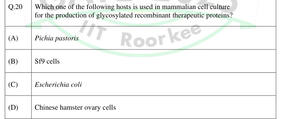
Which one of the following hosts is used in mammalian cell culture for the production of glycosylated recombinant therapeutic proteins?
- (A)
Pichia pastoris
- (B)
Sf9 cells
- (C)
Escherichia coli
- (D)
Chinese hamster ovary cells

GATE BT
DNA libraries, gene cloning and recombinant protein production
A DNA library is a collection of cloned fragments representing either a whole genome (a genomic library) or the mRNA population of a cell at one time (a cDNA library). A genomic library is made from partially digested or sheared DNA sized to the vector's capacity. A cDNA library is made from poly(A)+ mRNA by reverse transcription and second-strand synthesis. Library size follows N = ln(1-P)/ln(1-f). Libraries are screened by hybridization (sequence), antibodies (expressed protein) or functional complementation (restored phenotype), and genes are also isolated by homology PCR, chromosome walking, positional cloning and subtractive or differential methods. To produce a recombinant protein, the host (E. coli, yeast, insect or mammalian cells) and the construct (promoter, codons, fusion tags, secretion signals) are chosen to balance yield against correct folding and modification. Genes are disrupted at random by transposons, which leave target-site duplications, or at a chosen locus by homologous recombination with positive–negative selection, Cre–loxP or nuclease-induced breaks. DNA can be cloned without restriction sites by Gateway att-site recombination, λ Red recombineering or overlap-based assembly.
5 previous-year questions · 5 solved · 5 practice questions
A DNA library is a collection of cloned DNA fragments, each carried in a vector inside a host cell. Together the clones represent (ideally) the entire genome, which makes a genomic library, or the mRNA population of a cell or tissue at one time, which makes a cDNA library. Libraries are the starting material for isolating a gene and for surveying which genes are expressed.
Genomic library construction (in order)
Isolate high-molecular-weight genomic DNA.
Partially digest it with a frequently cutting restriction enzyme, or shear it randomly, to give overlapping fragments of clonable size. Sau3AI recognises 5′-↓GATC-3′ (a 4-bp site). The GATC overhang it leaves is compatible with the overhang of BamHI, 5′-G↓GATCC-3′, so the fragments can be ligated into a BamHI-cut vector. The hybrid junction is always recut by Sau3AI, but it re-forms a BamHI site only if the neighbouring bases happen to match. A complete digest is avoided: it gives a fixed set of non-overlapping fragments, some too large or too small to clone, and it splits any gene that contains a site.
Size-fractionate the fragments (e.g., by sucrose-gradient centrifugation or gel fractionation) to keep the range the chosen vector accepts (see the vector-capacity table).
Treat the vector with alkaline phosphatase to remove its 5′ phosphates so it cannot self-ligate, then ligate the fragments into it.
Package in vitro into phage heads (λ and cosmid vectors), or transform or electroporate (plasmid, BAC), or transform yeast (YAC). Plate to obtain individual clones.
Cloning vectors and approximate insert capacity (typical ranges; exact limits vary by vector)
Vector | Host | Approximate insert size |
|---|---|---|
Plasmid | E. coli | up to about 10 kb |
λ insertion vector (e.g., λgt10, λgt11) | E. coli | up to about 7–10 kb |
λ replacement vector | E. coli | about 9–23 kb |
Cosmid (packaged via cos sites; about 37–52 kb total between cos sites) | E. coli | about 35–45 kb |
Fosmid (single-copy F-plasmid based) | E. coli | about 35–45 kb |
BAC / PAC | E. coli | about 100–300 kb |
YAC | Yeast (S. cerevisiae) | about 100 kb to over 1 Mb |
cDNA library construction (in order)
Isolate total RNA and purify poly(A)+ mRNA on oligo(dT)-cellulose. This relies on the poly(A) tail of eukaryotic mRNA; bacterial mRNA generally lacks a stable poly(A) tail.
Make the first strand with reverse transcriptase primed by oligo(dT), or by random hexamers, which reach the 5′ regions of long mRNAs. The product is an mRNA–cDNA hybrid. Reverse transcriptase often stops before reaching the 5′ end of the mRNA, so many cDNAs are incomplete at the 5′ end.
Make the second strand by the replacement method (Gubler–Hoffman). RNase H nicks the mRNA strand, leaving short RNA primers; DNA polymerase I extends them by nick translation; E. coli DNA ligase seals the nicks; T4 DNA polymerase then makes the ends blunt. In the older self-priming method, a hairpin at the 3′ end of the first strand primes the second strand and S1 nuclease cuts the hairpin loop, which loses some sequence from the 5′ end of the mRNA.
Add ends for cloning. With linkers, first methylate internal EcoRI sites with EcoRI methylase, then ligate blunt EcoRI linkers and digest with EcoRI to create sticky ends. Adaptors already carry a sticky end, so no digestion is needed. Ligate into a vector: a plasmid, or a λ vector such as λgt10 (for hybridization screening) or λgt11 (an expression vector for antibody screening).
Transform or package and plate. Each clone's frequency in the library mirrors its mRNA's abundance at the time the RNA was isolated.
LIBRARY SIZE EQUATION (CLARKE–CARBON)
= number of independent clones required; = desired probability that a given sequence is present at least once. Genomic library: = average insert size / genome size, in the same units (bp). cDNA library: = the fraction of all mRNA molecules made up by the transcript of interest (its abundance). Assumes fragments are cloned at random and independently, with every fragment equally clonable. Always round up.
PROBABILITY THAT A GIVEN SEQUENCE IS PRESENT IN N CLONES
The same relation solved for . For small , , so , which slightly overestimates .
Genomic vs cDNA library
Feature | Genomic library | cDNA library |
|---|---|---|
Source material | Total genomic DNA | mRNA, copied into DNA by reverse transcriptase |
Introns, promoters, intergenic DNA | Present (the full gene structure) | Absent. Only the exon sequence of the mature mRNA: coding sequence plus 5′/3′ UTRs, often incomplete at the 5′ end |
Dependence on cell type | Essentially the same from any somatic cell of the organism (exceptions such as rearranged antigen-receptor loci in lymphocytes) | Specific to tissue, developmental stage and conditions |
Clone frequency | Each region represented roughly equally | Proportional to mRNA abundance, so rare transcripts need very large libraries |
Expressing a eukaryotic gene in E. coli | Not directly: bacteria cannot splice out introns | Possible once placed in a prokaryotic expression vector |
Typical use | Whole genes, regulatory regions, chromosome walking, genome mapping | Isolating expressed genes, expression cloning, producing recombinant proteins |
Typical vectors | Plasmid to very large-insert vectors (λ replacement, cosmid, BAC, YAC) | Plasmid or λ vectors (small-to-moderate inserts) |
EXAM TIP
A genomic library is made from a partial digest or randomly sheared DNA, so neighbouring clones overlap. A complete digest gives non-overlapping pieces that cannot be walked across, and it can split the gene of interest between two clones.
WORKED EXAMPLE
A genomic library is to be constructed for a genome of bp in cosmid vectors with an average insert size of 20 kb. What is the minimum number of clones needed for a 99% probability () that any given sequence is represented at least once?
Fraction of the genome per clone: .
.
.
Round up: .
Answer: 919 independent clones.
WORKED EXAMPLE
A particular mRNA makes up 1 in 50,000 mRNA molecules in a tissue. How many independent clones must a cDNA library from this tissue contain for a 99% probability that at least one clone carries this transcript?
For a cDNA library, is the abundance of the transcript: .
.
Round up: about clones.
Answer: About clones. Rare transcripts need far larger libraries than abundant ones.
Once a library exists, the clone carrying the gene of interest must be found among thousands to millions of clones. There are three broad screening strategies, and each tests a different property of the clone. Hybridization tests its sequence. Antibody screening tests the protein it produces; this works only if the insert is in an expression vector, in the correct orientation and reading frame. Functional complementation tests its biological activity.
Colony or plaque hybridization (sequence-based screening)
Replica-plate the colonies, or lift the plaques, onto a nitrocellulose or nylon membrane. Keep the master plate.
Lyse the cells or phage in situ, denature the released DNA to single strands, and fix it to the membrane by baking or UV cross-linking.
Hybridize with a labelled probe. A double-stranded probe must first be denatured. The probe can be a related gene, a degenerate oligonucleotide designed from partial protein sequence, or a heterologous probe from another species.
Wash at a stringency (temperature and salt) suited to the expected probe–target match. Higher temperature and lower salt mean higher stringency and less tolerance of mismatches.
Detect the positive signals (autoradiography or non-radioactive detection) and pick the matching colony or plaque from the master plate.
Antibody (immunological) screening, for expression libraries
The insert must be in an expression vector, in frame and in the correct orientation (e.g., a lacZ fusion in λgt11), so that the host makes the encoded protein.
Transfer the proteins expressed by the colonies or plaques onto a membrane.
Probe the membrane with an antibody specific to the protein of interest.
Detect the bound antibody with a labelled secondary antibody to find the positive clone.
Functional complementation screening
Introduce the library clones into a host strain carrying a specific mutation, for example an auxotroph or an enzyme-deficient strain.
Select or screen for restoration of the wild-type phenotype, such as growth on selective medium or restored enzyme activity.
A clone that complements the defect carries a functional copy of the corresponding gene.
The host must be able to express the clone. For a eukaryotic gene tested in a yeast or bacterial mutant, this generally means a cDNA library in an expression vector, because bacteria cannot splice out introns.
This isolates a gene purely by the function it restores, without any prior sequence or antibody information.
Screening methods compared
Method | What is detected | Prerequisite | Typical use |
|---|---|---|---|
Hybridization | Sequence similarity of DNA or RNA | A probe (homologous gene, degenerate oligonucleotide, or a gene from a related species) | Some sequence information is available |
Antibody screening | The expressed protein (epitope) | An expression vector with the insert in frame, and a specific antibody | The protein or an antibody is available but the sequence is not |
Functional complementation | Restoration of a phenotype | A suitable mutant host that can express the clone | Isolating a gene purely by its biological function |
EXAM TIP
Hybridization screening never requires the insert to be expressed. Expression is required only for antibody-based and functional screening of expression libraries.
Besides screening a library directly, several strategies are used to isolate a specific gene. The choice depends on what is already known about the gene: a related sequence, its map position, its differential expression, or only a phenotype.
Common gene isolation strategies
Homology-based cloning: design degenerate primers or probes from conserved regions of a known homologous protein, then use PCR or hybridization to pull out the gene.
RT-PCR and RACE: when part of the transcript sequence is known, amplify the cDNA with gene-specific primers. RACE (rapid amplification of cDNA ends) recovers the missing 5′ or 3′ ends.
Chromosome walking: use a probe from the end of one genomic clone to find the next, overlapping clone, and repeat to move along the chromosome toward a mapped gene. Chromosome jumping skips over large or unclonable stretches by circularising large fragments, so that DNA sequences far apart are brought together in one clone.
Positional (map-based) cloning: start from the gene's map location, defined by linked markers, and narrow it down by walking or jumping and by testing candidate genes. No prior knowledge of the gene product is needed.
Subtractive hybridization: hybridize cDNA from the tissue or condition of interest (the tester) with an excess of cDNA or mRNA from a reference sample (the driver), and remove the hybrids, which are the sequences common to both. This enriches for transcripts found only in, or increased in, the tester.
Differential screening: probe duplicate filters of the same library with labelled cDNA from two different samples, and pick clones that give a signal with only one of them.
Expression cloning: isolate the gene from an expression library purely by the activity or antigenicity of its protein product.
Which strategy for which situation
Known information | Suitable strategy |
|---|---|
A homologous gene or protein sequence exists | Homology-based cloning (degenerate primers or probes) |
Part of the transcript sequence is known | RT-PCR, with RACE for the missing ends |
The gene is genetically mapped but its function is unknown | Positional cloning, chromosome walking or jumping |
The gene is differentially expressed under some condition | Subtractive hybridization, differential screening |
Only the protein's activity, or an antibody against it, is available | Expression cloning (functional or antibody screening) |
Producing a recombinant protein means choosing a host and designing the construct so that the protein is made at a useful yield in a soluble, active and recoverable form. The choice depends on whether the protein needs post-translational modification, whether it is toxic to the host, and whether it tends to misfold.
Common expression host systems
Host | Growth and cost | Post-translational modification | Typical issues |
|---|---|---|---|
E. coli | Fast, cheap, often high yield | No eukaryotic-type N- or O-glycosylation. The cytoplasm is reducing, so disulfide bonds form poorly there; they form better in the periplasm or in engineered strains. | Many eukaryotic proteins form inclusion bodies; the protein is not glycosylated |
Yeast (e.g., Pichia pastoris, Saccharomyces cerevisiae) | Moderate cost; can secrete proteins into the medium | Glycosylates, but with high-mannose glycans unlike mammalian ones | S. cerevisiae in particular can hypermannosylate proteins, altering their properties |
Insect cells (baculovirus) | Moderate cost; slower than microbes | Glycosylates, mainly with simple (paucimannose) glycans that carry little sialic acid, unlike mammalian ones | Longer timelines than E. coli |
Mammalian cells (e.g., CHO) | Slow growth, costly media | Complex, mammalian-type glycosylation and folding | Highest cost and longest timelines; used when authentic modification is essential (e.g., therapeutic glycoproteins) |
Strategies to improve expression, solubility and recovery
Promoter and induction control: use a strong, regulated promoter (e.g., lac-derived or T7 systems induced with IPTG). Keep expression tightly off before induction if the protein is toxic to the host.
Codon optimization: replace codons that are rare in the host with codons the host prefers, to relieve translational stalling (or supply the rare tRNAs in the host).
Fusion tags: fuse an affinity tag (e.g., His-tag for metal-affinity purification, GST for glutathione affinity) or a solubility partner (e.g., MBP, thioredoxin, SUMO) in frame. These aid detection and one-step purification, and often improve solubility. Include a specific protease cleavage site if the tag must be removed.
Secretion signals: add a signal peptide to export the protein to the periplasm or the medium. The oxidizing periplasm favours disulfide bonds, and secretion leaves fewer host proteins to purify away.
Controlling inclusion bodies: lower the induction temperature and inducer concentration, co-express molecular chaperones, or use a solubility tag. If inclusion bodies still form, recover the protein by solubilizing it in a denaturant (e.g., urea or guanidine hydrochloride) and refolding it under controlled conditions.
Host and strain choice: match the host to the protein's modification needs (a mammalian or insect system, not E. coli, when function depends on authentic glycosylation). Protease-deficient strains reduce degradation.
EXAM TIP
Inclusion bodies are dense aggregates of usually inactive protein. They do not mean the gene failed to express: they often form because the protein is made faster than it can fold. The usual remedies are to reduce the rate of expression so folding can keep up, or to solubilize and refold the aggregates. Inclusion bodies are easy to isolate by centrifugation and resist proteolysis, which can make them useful for toxic or protease-sensitive proteins.
WORKED EXAMPLE
A eukaryotic enzyme is expressed at a very high level in E. coli from a strong inducible promoter, but almost all of it is recovered as inactive protein in the insoluble pellet after lysis. Give three strategies to try before switching host, in a sensible order, and justify each briefly.
Diagnosis: very high expression with an inactive, insoluble product is the typical sign of inclusion bodies. The protein is being made faster than it folds; this is not a cloning or transcription failure.
Strategy 1: lower the induction temperature (e.g., 37 °C to about 16–20 °C) and the inducer concentration. This slows synthesis and gives each chain more time to fold before it aggregates.
Strategy 2: fuse an in-frame solubility tag (e.g., MBP or thioredoxin), with a protease site for later removal. Such tags often reduce aggregation of the partner protein.
Strategy 3: co-express molecular chaperones (e.g., GroEL/GroES). They help nascent chains fold and can reduce aggregation.
If these fail, solubilize and refold the inclusion bodies, or move to another host (yeast, insect or mammalian cells), which costs more time and money.
Answer: (1) Lower the induction temperature and inducer concentration; (2) add a solubility-enhancing fusion tag; (3) co-express chaperones. Only then refold, or change host.
Genes can be disrupted or modified in two complementary ways. Transposon mutagenesis inserts DNA at essentially random sites across the genome. Gene targeting changes a chosen locus, by homologous recombination with flanking homology, often helped by site-specific recombinases or by a double-strand break made by a nuclease.
Transposable elements: core facts
Transposons are DNA segments that move from one site in a genome to another. DNA transposons are moved by a transposase, usually encoded by the element itself. Transposition does not need sequence homology between the element and its target site.
Insertion sequences (IS elements) are the simplest bacterial transposons: a transposase gene flanked by short terminal inverted repeats.
Composite transposons (e.g., Tn5, Tn10) carry a central region, often an antibiotic-resistance gene, flanked by two IS elements.
Insertion creates a target-site duplication: a few base pairs of host DNA, repeated directly on both sides of the element, because the transposase cuts the target with a staggered cut.
Conservative (cut-and-paste) transposition excises the element and reinserts it elsewhere (e.g., Tn5, Tn10). Replicative transposition copies the element and leaves the original in place (e.g., Tn3, through a cointegrate that a resolvase separates).
Retrotransposons move through an RNA intermediate that is reverse-transcribed into DNA and inserted (copy-and-paste). LTR retrotransposons are flanked by long terminal repeats in direct orientation.
Uses in cloning and mutagenesis: transposon tagging, where an insertion inactivates a gene and its marker lets the gene be recovered (e.g., the flanking DNA by inverse PCR or plasmid rescue); P-element transformation of Drosophila; engineered vertebrate transposon vectors (Sleeping Beauty, piggyBac); and Tn7 transposition used to build recombinant baculovirus shuttle vectors (bacmids).
Gene targeting by homologous recombination (a typical knockout)
Design a targeting construct with two homology arms matching the sequence on either side of the gene. Between the arms, a positive selectable marker (e.g., neo, which confers G418 resistance) replaces part or all of the gene.
Optionally, place a negative selectable marker (e.g., HSV-tk) outside one homology arm. Correct homologous recombination exchanges only the DNA between the arms, so the outside marker is lost. Random (non-homologous) integration usually inserts the whole construct, so the marker is normally retained.
Introduce the linearized construct into the target cells, e.g., mouse embryonic stem cells, by electroporation.
Apply positive selection (G418, which keeps any stable integrant) and negative selection (ganciclovir, which is toxic to cells expressing HSV-tk) together. This enriches for correctly targeted clones.
Confirm correct targeting in the surviving clones by PCR or Southern blot. In mammalian cells, homologous recombination is much rarer than random integration, so enrichment is not proof of targeting. In yeast, homologous recombination is efficient.
Site-specific recombinases and targeted nucleases
Cre–loxP: Cre recombinase, from bacteriophage P1, recombines two 34-bp loxP sites. Each site is two 13-bp inverted repeats around an asymmetric 8-bp spacer. Two loxP sites in the same orientation on one molecule lead to excision of the DNA between them, leaving one loxP. Sites in opposite orientations lead to inversion. Flanking an exon with loxP sites ('floxing') and expressing Cre only in one tissue or at one time gives a conditional knockout.
FLP–FRT: the yeast FLP recombinase acting on FRT sites works the same way; it is often used to remove selection markers.
Nuclease-assisted targeting: a double-strand break made at the chosen locus greatly raises the rate of targeted change. In CRISPR–Cas9, the Cas9 nuclease is guided by a guide RNA whose roughly 20-nt spacer pairs with the target, next to a PAM (5′-NGG-3′ for the commonly used Streptococcus pyogenes Cas9). Repair by non-homologous end joining (NHEJ) leaves small insertions or deletions, often frameshifts, which gives a knockout. Repair by homology-directed repair (HDR) copies a supplied donor template and gives a precise edit or insertion. Zinc-finger nucleases and TALENs make targeted breaks with protein-based DNA recognition.
Transposon mutagenesis vs targeted knockout
Feature | Transposon mutagenesis | Targeted knockout by homologous recombination |
|---|---|---|
Site of insertion or change | Essentially random across the genome | A predetermined locus |
Typical use | Large-scale mutagenesis screens and gene discovery | Studying a specific, already identified gene |
Efficiency at a chosen locus | Low: many random insertions must be screened to hit one gene | Needs enrichment (positive–negative selection) or a nuclease-induced break, because random integration competes with it |
Role of the marker | Tags the disrupted gene, so it can be identified afterwards | Placed deliberately by the researcher; the gene is already known |
EXAM TIP
Positive selection alone cannot tell random integration from true homologous recombination, because both integrate the marker. Negative selection against a marker placed outside the homology arms enriches for correctly targeted clones, because random integrants usually keep that marker.
Several methods move or assemble DNA without depending on restriction sites. Site-specific recombination cloning (the Gateway system) and homologous-recombination cloning (λ Red recombineering, yeast gap repair) use recombination. Overlap-based assembly (Gibson assembly) joins fragments through designed overlapping sequence and uses no recombinase.
Gateway cloning (site-specific recombination at att sites of phage λ)
The gene of interest is flanked by attB sites, by PCR or an earlier cloning step.
BP reaction: BP Clonase (λ integrase, Int, plus E. coli integration host factor, IHF) recombines the attB sites with the attP sites of a donor vector. The product is an entry clone flanked by attL sites, plus a by-product carrying attR sites.
LR reaction: LR Clonase (Int, λ excisionase Xis, and IHF) recombines the attL sites of the entry clone with the attR sites of a chosen destination vector. The product is an expression clone with attB sites regenerated, plus a by-product with attP sites.
The donor and destination vectors carry the toxic ccdB gene between their att sites. Unreacted vectors therefore kill standard E. coli, while recombined clones, which have lost ccdB, survive.
Recombination is directional and conservative, so one entry clone can be moved into many destination vectors (different promoters, tags or hosts) without re-cloning the gene.
Homologous-recombination cloning
λ Red recombineering in E. coli: three phage λ proteins carry out recombination. Exo is a 5′→3′ exonuclease that exposes single-stranded 3′ ends. Beta binds single-stranded DNA and anneals it to its complementary target. Gam inhibits the host RecBCD nuclease, protecting the linear DNA. Short homology arms, typically about 35–50 bp added on PCR primers, are enough to insert, delete or modify sequence in plasmids, BACs or the chromosome.
Yeast gap-repair cloning: a linearized vector and an insert whose ends match the vector ends are co-transformed into S. cerevisiae. The yeast's efficient homologous recombination joins them into a circular plasmid.
Gibson assembly (isothermal, overlap-based, seamless)
Design the fragments (inserts and linearized vector) so that the ends to be joined share overlapping sequence, typically about 20–40 bp.
Mix the fragments with one enzyme cocktail containing a 5′→3′ exonuclease, a DNA polymerase and a DNA ligase, and incubate at a single temperature, commonly 50 °C.
The exonuclease chews back the 5′ ends, exposing single-stranded 3′ overhangs at the overlaps, which anneal to their partner fragments.
The polymerase fills the gaps and the ligase seals the nicks. Several fragments are joined in their designed order in one reaction, with no restriction-site scar.
Comparing cloning approaches
Method | Basis of joining | Scar or extra sequence | Best suited for |
|---|---|---|---|
Restriction–ligation | Compatible sticky or blunt ends made by restriction enzymes | The restriction site usually remains at the junction | Simple single-insert cloning when a suitable unique site exists |
Gateway | Site-specific recombination between att sites (Int/IHF, with Xis for LR) | A short att sequence remains at each junction | Moving one gene into many different destination vectors |
λ Red recombineering / yeast gap repair | Homologous recombination through short homology arms | None beyond the designed sequence | Editing large constructs (BACs, chromosomes) or joining fragments inside the host |
Gibson assembly | Designed overlaps, with an exonuclease, polymerase and ligase working in vitro | Seamless when designed to be | Joining several fragments in one step, independent of restriction sites |
EXAM TIP
Gateway cloning depends on att-site recombination by λ Int, IHF and Xis, not on restriction enzymes or designed overlaps, so it works even when no unique restriction site exists. Gibson assembly depends only on designed overlaps between fragment ends and involves no recombinase or att site. Recombineering depends on homologous recombination through short homology arms.
PREVIOUS-YEAR QUESTIONS
Try each question first, then open the answer. We placed each question under a topic ourselves; the exam paper does not label topics.
See these papers in full: GATE BT 2025 · GATE BT 2020
Which one of the following hosts is used in mammalian cell culture for the production of glycosylated recombinant therapeutic proteins?
Pichia pastoris
Sf9 cells
Escherichia coli
Chinese hamster ovary cells
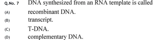
DNA synthesized from an RNA template is called
recombinant DNA.
transcript.
T-DNA.
complementary DNA.
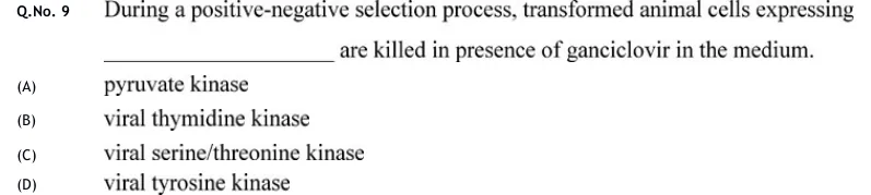
During a positive-negative selection process, transformed animal cells expressing ________ are killed in presence of ganciclovir in the medium.
pyruvate kinase
viral thymidine kinase
viral serine/threonine kinase
viral tyrosine kinase
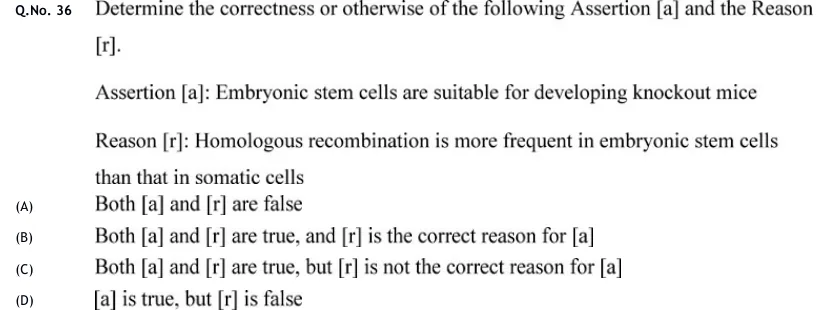
Determine the correctness or otherwise of the following Assertion [a] and the Reason [r].
Assertion [a]: Embryonic stem cells are suitable for developing knockout mice
Reason [r]: Homologous recombination is more frequent in embryonic stem cells than that in somatic cells
Both [a] and [r] are false
Both [a] and [r] are true, and [r] is the correct reason for [a]
Both [a] and [r] are true, but [r] is not the correct reason for [a]
[a] is true, but [r] is false
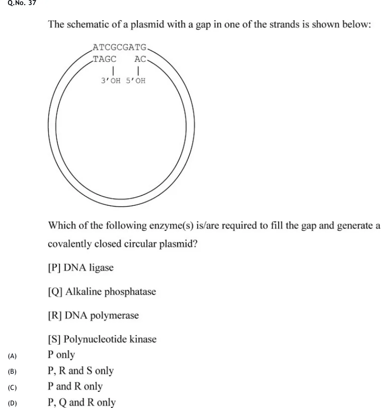
The schematic of a plasmid with a gap in one of the strands is shown below: [circular double-stranded plasmid diagram]. Top strand (continuous, outer): 5'-ATCGCGATG-3'. Bottom strand (broken, shown below it): left fragment 'TAGC' ending in a vertical line down to '3'OH', then a gap, then right fragment 'AC' with a vertical line down to '5'OH'. So the bottom strand is interrupted: one piece ends 3'-OH (after TAGC), and the next piece begins 5'-OH (before AC), with several bases of the complementary strand missing between them (a gap), opposite the continuous top strand ATCGCGATG. Which of the following enzyme(s) is/are required to fill the gap and generate a covalently closed circular plasmid? [P] DNA ligase [Q] Alkaline phosphatase [R] DNA polymerase [S] Polynucleotide kinase
 Tap to open full size
A circular plasmid drawn as two concentric circles (double-stranded DNA, most of the circle shown as plain double lines representing the rest of the plasmid). At one point on the circle, the sequence is spelled out: the outer/top strand reads 5'-ATCGCGATG-3' continuously (no break). Directly below it, aligned by base-pairing, the bottom strand is shown broken into two fragments with a gap between them: the left fragment reads 'TAGC' and has a line dropping down to the label '3'OH' (marking its 3' terminus); after a gap of missing bases, the right fragment reads 'AC' and has a line dropping down to the label '5'OH' (marking its 5' terminus, explicitly written as 5'-OH rather than 5'-phosphate). This depicts a plasmid strand with both a gap (missing nucleotides) and, once the gap is filled, a nick whose 5' end lacks a phosphate group.
P only
P, R and S only
P and R only
P, Q and R only
PRACTICE
In the replacement (RNase H–based) method of cDNA second-strand synthesis, RNase H nicks the mRNA strand of the mRNA–cDNA hybrid, leaving short RNA fragments that act as primers. Which enzyme then synthesizes the second (sense) cDNA strand?
Reverse transcriptase
DNA polymerase I
T4 DNA ligase
Terminal transferase
Answer: (B)
Reverse transcriptase makes the first-strand cDNA on the mRNA template, giving an mRNA–cDNA hybrid.
RNase H nicks and partially degrades the mRNA strand, leaving short RNA fragments annealed to the first strand. These act as primers.
DNA polymerase I extends these primers by nick translation, replacing the RNA fragments with DNA and making the second strand.
DNA ligase seals the remaining nicks. The ends are then made blunt with T4 DNA polymerase before linkers or adaptors are added.
The enzyme that synthesizes the second strand is therefore DNA polymerase I.
A genomic DNA library is being constructed for a genome of bp, with an average insert size of 15 kb. Using , find the minimum number of independent clones needed for a probability that any given sequence is represented at least once. Round up to the next whole number.
Answer: (accepted range )
Fraction of the genome per clone: .
.
.
A minimum number of clones is rounded up: .
Which of the following statements about screening a genomic DNA library by colony or plaque hybridization with a labelled nucleic-acid probe are correct?
A double-stranded DNA probe must be labelled and denatured to single strands before use
Colonies or plaques are transferred to a membrane and lysed in situ before hybridization
The technique requires the cloned insert to be expressed as a functional protein
The stringency of hybridization and washing controls how many mismatches between probe and target are tolerated
Answer: (A), (B), (D)
A: True. A double-stranded probe must be labelled and denatured into single strands so that it can base-pair with the denatured library DNA on the membrane.
B: True. Colonies or plaques are lifted onto a nitrocellulose or nylon membrane, lysed in situ, and their DNA is denatured and fixed before hybridization.
C: False. Hybridization detects sequence complementarity only. It does not need the insert to be expressed as a protein; that requirement applies to antibody or functional screening of expression libraries.
D: True. The temperature and salt concentration of hybridization and washing set how many mismatches between probe and target are tolerated, and so control specificity.
In Gateway cloning, which enzyme mixture transfers the gene of interest from an entry clone (flanked by attL sites) into a destination vector (carrying attR sites) to give an expression clone?
BP Clonase
LR Clonase
T4 DNA ligase
A restriction endonuclease alone
Answer: (B)
Gateway cloning uses site-specific recombination between att sites, catalysed by enzymes from phage λ and E. coli.
The BP reaction (BP Clonase: Int + IHF; attB × attP) moves a PCR product into a donor vector to create the entry clone, which has attL sites.
The LR reaction (LR Clonase: Int + Xis + IHF; attL × attR) moves the insert from the entry clone into a destination vector, creating an expression clone with attB sites. Unreacted destination vector still carries ccdB and is selected against.
Entry clone to destination vector is therefore the LR reaction, catalysed by LR Clonase.
A knockout construct for homologous recombination in mouse embryonic stem cells has a neomycin-resistance (neo) cassette between two homology arms, and a herpes simplex virus thymidine kinase (HSV-tk) cassette outside one homology arm (a positive–negative selection design). After electroporation, the cells are grown in G418 plus ganciclovir. What does this double selection enrich for?
Random integrants only
Correctly targeted (homologous recombination) clones, by killing random integrants that retain HSV-tk
Untransfected cells only
Cells that have completely lost the neo cassette
Answer: (B)
G418 kills cells that have not stably integrated the neo cassette, so every survivor carries neo, whether it was targeted or integrated at random.
Random (non-homologous) integration usually inserts the whole linear construct, including the outside HSV-tk cassette. HSV-tk converts ganciclovir into a toxic nucleotide analogue, so most random integrants die.
Correct homologous recombination exchanges only the DNA between the homology arms, so the outside HSV-tk cassette is lost and correctly targeted cells survive ganciclovir.
Together the two drugs enrich for correctly targeted clones. The survivors are still confirmed by PCR or Southern blot, because the enrichment is not absolute.
OFFICIAL GATE 2027 SYLLABUS WORDING
Copied from the GATE 2027 syllabus; see all 2027 syllabi.
GATE BT
cDNA and genomic DNA library Gene isolation and cloning, strategies for production of recombinant proteins; Transposons and gene targeting, Recombination-based gene cloning
Preparing in Kolkata? IAE Academy's course for GATE BT.