IAE Academy

GATE BT

Genes and chromosomes, mutation and DNA repair

Prokaryotic genes are often grouped in operons and read from polycistronic mRNAs with a Shine–Dalgarno site before each start codon. Most eukaryotic genes are monocistronic and split into exons and introns (GU–AG rule), and their transcripts are capped, spliced and polyadenylated. Eukaryotic DNA is packaged into chromatin, beginning with nucleosome core particles of about 147 bp of DNA in 1.65 left-handed turns around a histone octamer. Centromeres and telomeres (with telomerase) maintain chromosome integrity. A mutation is a heritable sequence change: a transition or transversion, silent, missense or nonsense, or a frameshift. Mutations arise spontaneously or from mutagens (base analogues, deaminating, hydroxylating, alkylating and intercalating agents, UV, ionizing radiation). The Ames test screens chemicals for mutagenicity. Each class of DNA damage is handled by a characteristic pathway: direct reversal, BER, NER, MMR, or HR and NHEJ for double-strand breaks. Translesion synthesis tolerates damage rather than repairing it.

9 previous-year questions · 9 solved · 6 practice questions

Molecular structure of genes: prokaryotic and eukaryotic

A gene is the segment of DNA that carries the information (and regulatory signals) needed to produce a functional RNA or protein product. Its physical organisation differs sharply between prokaryotes and eukaryotes, and this difference underlies how mutations and their downstream effects are interpreted.

EXAM TIP

Convention: nucleic acid sequences are written 5' to 3'. RNA polymerase reads the template (antisense) strand 3' to 5' and synthesises the mRNA 5' to 3'. The mRNA therefore has the same sequence as the coding (sense) strand, with U in place of T. Codons, including the stop codons UAA, UAG and UGA, are quoted as mRNA triplets read 5' to 3'. Gene symbols are written plainly (lacZ, uvrB).

In prokaryotes, functionally related genes are often organised into operons. A single promoter (with an operator, where one is present) drives one polycistronic mRNA that encodes several proteins, e.g., the lac operon (lacZ, lacY, lacA). Each open reading frame (ORF) in such an mRNA usually has its own Shine–Dalgarno sequence (ribosome-binding site). This is a purine-rich stretch a few nucleotides upstream of the start codon that base-pairs with the 3' end of 16S rRNA, so each cistron can be initiated separately. Bacterial protein-coding genes almost never contain introns.

In eukaryotes, each protein-coding gene is usually transcribed as its own monocistronic transcript. In multicellular eukaryotes most such genes are split: coding exons are interrupted by non-coding introns, although some genes, such as most replication-dependent histone genes, have none. The primary transcript (pre-mRNA) is processed in the nucleus. It receives a 5' 7-methylguanosine cap, the spliceosome removes the introns and joins the exons, and the 3' end is cleaved and polyadenylated. The mature mRNA is then exported to the cytoplasm. Much of this processing happens while transcription is still in progress (co-transcriptionally).

Almost all nuclear pre-mRNA introns begin with GU at the 5' splice site and end with AG at the 3' splice site (the GU–AG rule). A branch-point adenosine lies a short distance upstream of the 3' splice site. Splicing proceeds by two transesterification reactions and releases the intron as a lariat. A mutation in a splice site or branch point can cause exon skipping or intron retention even though no codon is changed.

Order of elements in a typical eukaryotic protein-coding gene and its transcript (5' to 3' along the coding strand)

  1. Enhancers and other distal regulatory elements. These can lie upstream, downstream or within introns, often far from the promoter, and work largely regardless of orientation.

  2. Core promoter, around and just upstream of the transcription start site: e.g., a TATA box about 25–30 bp upstream or an initiator (Inr) at the start site. Many promoters lack a TATA box.

  3. Exon 1, beginning at the transcription start site. Its first part is the 5' untranslated region (5' UTR). The start codon (AUG in the mRNA) lies downstream of it, in exon 1 or a later exon.

  4. Intron 1, exon 2, intron 2, … up to the last exon. Introns are removed and exons joined in the mature mRNA.

  5. Last exon: contains the stop codon, followed by the 3' UTR, which carries the polyadenylation signal (AAUAAA in the transcript). The transcript is cleaved a short distance downstream of this signal and a poly(A) tail is added.

  6. Transcription termination region, beyond the cleavage site.

Prokaryotic vs eukaryotic gene organisation

Feature

Prokaryotic gene

Eukaryotic gene

Introns

Absent from almost all bacterial protein-coding genes

Present in most genes of multicellular eukaryotes; removed by splicing

Transcript

Often polycistronic (operon)

Usually monocistronic

Promoter recognition

Sigma factor of the RNA polymerase holoenzyme recognises the −10 and −35 elements

RNA polymerase II with general transcription factors (e.g., TFIID binds the TATA box)

Ribosome binding

Shine–Dalgarno sequence upstream of each start codon

Small ribosomal subunit binds the 5' cap and scans to the first AUG in a favourable context (Kozak sequence)

Transcription and translation

Coupled: translation can begin on an mRNA that is still being transcribed

Separated in space and time: transcription and processing in the nucleus, translation in the cytoplasm

mRNA processing

Little or none

5' capping, splicing, 3' cleavage and polyadenylation

Chromosome and chromatin organisation

Eukaryotic nuclear DNA is packaged with proteins into chromatin. Histones are small basic proteins rich in lysine and arginine, and their positive charges bind the negatively charged phosphate backbone. The repeating unit is the nucleosome. Its core particle is a histone octamer: an (H3–H4)₂ tetramer flanked by two H2A–H2B dimers, i.e., two copies each of H2A, H2B, H3 and H4. About 147 bp of DNA (older value 146 bp) is wrapped around the octamer in about 1.65 left-handed superhelical turns. Adjacent core particles are joined by linker DNA of variable length. The linker histone H1 binds where DNA enters and leaves the core particle and helps compact the 'beads-on-a-string' fibre into higher-order structure.

Limited digestion of chromatin with micrococcal nuclease cuts the exposed linker DNA and gives a ladder of DNA fragments in multiples of the nucleosome repeat length. That length is about 200 bp in many cells and varies between cell types and species. More extensive digestion trims each particle down to the protected core DNA.

NUCLEOSOME REPEAT LENGTH

WORKED EXAMPLE

In a certain cell type the nucleosome repeat length is 195 bp. Taking the DNA of one core particle as 147 bp, how much linker DNA lies between adjacent core particles?

Solution
  1. Repeat length = core-particle DNA + linker DNA.

  2. Linker DNA = 195 − 147 = 48 bp.

Answer: 48 bp

Levels of chromatin packaging (increasing compaction)

  1. Naked double-helical DNA (about 2 nm wide)

  2. 11 nm 'beads-on-a-string' fibre: nucleosome core particles joined by linker DNA

  3. 30 nm fibre: a textbook model of nucleosomes coiled with the help of H1. Whether a regular 30 nm fibre exists in most living interphase nuclei is debated.

  4. Looped domains anchored to a non-histone protein scaffold

  5. Condensed metaphase chromosome (maximal compaction, visible by light microscopy)

Chromatin state is regulated by covalent histone modifications, mainly on the N-terminal tails. Acetylation of lysines by histone acetyltransferases neutralises their positive charge and is generally associated with open, transcriptionally active chromatin. Deacetylation, and marks such as trimethylation of lysine 9 on histone H3 (H3K9me3), are associated with condensed, silent heterochromatin. In vertebrates, methylation of cytosine at CpG sites (5-methylcytosine) in promoters is associated with gene silencing.

Heterochromatin vs euchromatin

Feature

Heterochromatin

Euchromatin

Compaction

Highly condensed

Relatively decondensed

Transcriptional activity

Largely silent

Active or competent for transcription

Staining (interphase nuclei)

Stains darkly

Stains lightly

Replication timing

Late in S phase

Earlier in S phase

Typical histone marks

Hypoacetylated; H3K9 methylation

Acetylated histones

Types and examples

Constitutive: condensed in all cells, e.g., pericentromeric satellite repeats. Facultative: condensed only in some cells, e.g., the inactive X (Barr body) in female mammals

Bulk of the actively expressed genome

The centromere is the primary constriction of a chromosome. The kinetochore assembles there to attach spindle microtubules, and cohesion between sister chromatids persists there until anaphase. Its chromatin contains a centromere-specific histone H3 variant (CENP-A), and it is usually flanked by heterochromatin.

Telomeres are tandem repeats at chromosome ends (TTAGGG in vertebrates). The G-rich strand runs 5' to 3' towards the end and finishes as a single-stranded 3' overhang. They are bound by a protective protein complex (shelterin in mammals), which stops the ends from being recognised as double-strand breaks. Telomeres also buffer the sequence lost through the end-replication problem: at the very end of the lagging strand the last RNA primer is removed, and the gap cannot be filled because DNA polymerase needs a 3'-OH primer. Telomerase is a ribonucleoprotein reverse transcriptase that carries its own RNA template and extends the 3' G-rich strand. It is active in germ cells, many stem cells and most human cancers, but low or absent in most human somatic cells. Their telomeres therefore shorten with successive divisions, which contributes to replicative senescence.

Types of mutations

A mutation is a heritable change in the nucleotide sequence of DNA. Mutations are classified by their molecular nature (base substitutions, insertions and deletions, larger rearrangements) and, in coding sequence, by their effect on the encoded protein. They are spontaneous when they come from replication errors, tautomeric shifts, or spontaneous depurination and deamination, and induced when they are caused by mutagens.

Base substitutions: transitions and transversions

Type

Definition

Examples

Transition

A purine is replaced by the other purine, or a pyrimidine by the other pyrimidine

A·T → G·C; G·C → A·T

Transversion

A purine is replaced by a pyrimidine, or a pyrimidine by a purine

G·C → T·A; A·T → C·G; A·T → T·A; G·C → C·G

Count

Each base has one possible transition and two possible transversions (of the 12 possible single-base changes, 4 are transitions and 8 are transversions)

e.g., A → G is a transition; A → C and A → T are transversions

Point mutations in coding sequence: effect on the encoded protein

Type

Definition

Effect on protein

Silent (synonymous)

The new codon specifies the same amino acid (codon degeneracy); most often a change at the third (wobble) position

No change in amino acid sequence

Missense

The new codon specifies a different amino acid; conservative (chemically similar) or non-conservative

Single amino acid substitution; the effect ranges from negligible to loss of function, depending on position and chemistry

Nonsense

A sense codon becomes a stop codon (UAA, UAG or UGA in the mRNA, read 5' to 3')

Premature termination; truncated, usually non-functional protein

Frameshift mutations

  1. Caused by inserting or deleting a number of nucleotides that is NOT a multiple of three

  2. Shifts the triplet reading frame for every codon downstream of the site

  3. Alters all downstream amino acids and very often creates a premature stop codon in the new frame

  4. An insertion or deletion that IS a multiple of three adds or removes whole codons without shifting the frame (an in-frame indel). It can still create a stop codon at the junction.

Other mutation classes

  1. Tautomeric shifts: rare tautomers (imino or enol forms) mispair during replication, e.g., enol T pairs with G. This produces transitions.

  2. Forward vs reverse mutation: a reversion restores the wild-type phenotype, either by restoring the original sequence or by a suppressor mutation. An intragenic suppressor is a second change in the same gene, e.g., a nearby insertion that restores the frame after a deletion. An intergenic suppressor lies in another gene, e.g., a suppressor tRNA that reads a stop codon.

  3. Loss-of-function mutations are usually recessive; gain-of-function mutations are often dominant.

  4. Conditional mutations show their phenotype only under restrictive conditions, e.g., temperature-sensitive alleles.

EXAM TIP

'Synonymous' and 'silent' both mean no change in the amino acid sequence. A synonymous change is not always completely neutral, since some affect splicing, mRNA stability or translation speed. For classifying mutations, treat silent as no change in the protein's amino acid sequence.

Mutagens and their mechanisms; the Ames test

Major classes of mutagens

Class

Example(s)

Mechanism

Typical mutation caused

Base analogues

5-bromouracil (5-BU); 2-aminopurine (2-AP)

5-BU replaces T. Its common keto form pairs with A; its rarer enol (or ionised) form pairs with G. 2-AP replaces A and pairs with T, but occasionally with C. The change becomes fixed only after further rounds of replication.

Transitions (A·T ↔ G·C, in both directions)

Deaminating agent

Nitrous acid (HNO₂)

Deaminates C → U (pairs with A), A → hypoxanthine (pairs with C) and G → xanthine (still pairs with C, so usually not mutagenic)

Transitions: G·C → A·T (from C deamination) and A·T → G·C (from A deamination)

Hydroxylating agent

Hydroxylamine (NH₂OH)

Hydroxylates the amino group of cytosine; the product pairs with A

Transitions in one direction only: G·C → A·T

Alkylating agents

Ethyl methanesulfonate (EMS), nitrosoguanidine, sulfur mustard

Add alkyl groups to bases. O6-ethylguanine (from EMS) pairs with T. Alkylation at N7 of guanine weakens the glycosidic bond and promotes depurination. Bifunctional agents such as mustards form cross-links.

Mainly G·C → A·T transitions (EMS); also transversions, deletions and strand breaks via depurination and cross-links

Intercalating agents

Acridine orange, proflavine, ethidium bromide

Flat ring systems slip between stacked base pairs and stretch the helix, so a base is added or skipped during replication

Insertion or deletion of one or a few base pairs, i.e., frameshifts

UV radiation

UV-C (about 254 nm, close to the DNA absorption maximum near 260 nm)

Covalently links adjacent pyrimidines on the same strand, forming cyclobutane pyrimidine dimers (CPDs; most often T–T) and 6-4 photoproducts

Helix-distorting lesions that block polymerases. Mutations arise when error-prone translesion synthesis bypasses them; C → T transitions at dipyrimidine sites are characteristic.

Ionizing radiation

X-rays, gamma rays

Direct ionisation and free radicals (especially hydroxyl radicals from water) cause base damage and single- and double-strand breaks

Deletions, chromosome breaks and rearrangements

Ames test: standard procedure

  1. Tester strains are histidine auxotrophs (his⁻) of Salmonella enterica serovar Typhimurium (written S. typhimurium in older literature). Each carries a known mutation in a histidine-biosynthesis gene and cannot grow without histidine.

  2. Different strains carry different his mutations, so they detect different classes of mutagen. A strain with a base-substitution mutation (e.g., TA100) is reverted by base-substituting mutagens; a strain with a frameshift mutation (e.g., TA98) is reverted by frameshift mutagens.

  3. The strains are made more sensitive. A uvrB deletion removes nucleotide excision repair. An rfa mutation weakens the lipopolysaccharide coat and lets large molecules in. Several strains also carry the plasmid pKM101, which has error-prone repair genes.

  4. Bacteria are plated on minimal glucose agar with the test chemical at several doses. A trace of histidine is added so that every cell can divide a few times, which is needed for mutations to become fixed, but not enough to form visible colonies.

  5. Parallel plates include rat liver S9 fraction: the supernatant of liver homogenate centrifuged at 9000 g, which contains microsomal enzymes such as cytochrome P450. It activates pro-mutagens that need metabolic conversion.

  6. After incubation, only his⁺ revertants form colonies. A reproducible, dose-dependent increase over the spontaneous (solvent-control) revertant count indicates that the chemical is mutagenic.

EXAM TIP

Some chemicals (pro-mutagens) are harmless until liver enzymes convert them into a reactive, DNA-damaging form, so the Ames test is run both with and without S9. A positive result shows mutagenicity in bacteria and is used as a screen for possible carcinogens. It is not a direct test of carcinogenicity.

Types of DNA damage

Common types of spontaneous and induced DNA damage

Damage type

Cause

Description

Depurination

Spontaneous hydrolysis of the N-glycosidic bond (faster at high temperature and low pH; promoted by N7-alkylation of guanine)

Loss of a purine (A or G), leaving an apurinic (AP, abasic) site

Deamination

Spontaneous, or induced by nitrous acid

C → U (pairs with A). 5-methylcytosine → T, a normal base, leaving a T·G mismatch; this makes methylated CpG sites mutational hotspots. A → hypoxanthine (pairs with C). G → xanthine.

Pyrimidine dimers

UV light

Cyclobutane pyrimidine dimers and 6-4 photoproducts between adjacent pyrimidines on the same strand; they distort the helix and block replication and transcription

Alkylation adducts

Alkylating agents (EMS, nitrosoguanidine, mustards)

Alkyl groups added to bases, e.g., O6-methylguanine (pairs with T); N7-methylguanine and N3-methyladenine

Oxidative damage

Reactive oxygen species (ROS)

Oxidised bases such as 8-oxoguanine, which can pair with A; unrepaired, it gives G·C → T·A transversions

Cross-links

Bifunctional alkylating agents (mustards), cisplatin, psoralen with UVA

Covalent links within one strand (intrastrand) or between the two strands (interstrand); interstrand cross-links block strand separation

Strand breaks

Ionizing radiation, ROS, some chemicals, collapsed replication forks

Single-strand breaks (SSBs) or double-strand breaks (DSBs) in the sugar–phosphate backbone

DNA repair pathways

Three successive lines of defence keep replication accurate: base selection by the DNA polymerase, the polymerase's own 3' to 5' exonuclease proofreading, and then mismatch repair of errors that escape proofreading. Chemical damage to DNA is handled by the pathways below.

Overview of major repair and damage-tolerance pathways

Pathway

Damage handled

Key features

Direct reversal

O6-alkylguanine; cyclobutane pyrimidine dimers (photoreactivation)

The damage is reversed chemically without removing nucleotides. O6-methylguanine–DNA methyltransferase (MGMT; Ada in E. coli) transfers the alkyl group to one of its own cysteines and is permanently inactivated: a 'suicide' enzyme, one molecule per lesion. DNA photolyase uses visible (blue) or near-UV light and a reduced FAD cofactor to split CPDs. Photolyase is absent in placental mammals, including humans.

Base excision repair (BER)

Small, non-distorting base lesions: uracil, deaminated, oxidised or alkylated bases; AP sites

A lesion-specific DNA glycosylase cuts the N-glycosidic bond (e.g., uracil–DNA glycosylase) and leaves an AP site. AP endonuclease nicks the backbone on the 5' side of the AP site. The gap is filled (in mammals usually a single nucleotide, by DNA polymerase β) and DNA ligase seals it.

Nucleotide excision repair (NER)

Bulky, helix-distorting lesions: pyrimidine dimers, 6-4 photoproducts, large chemical adducts

Recognises the helix distortion rather than the specific chemistry, cuts the damaged strand on both sides of the lesion and removes an oligonucleotide (about 12–13 nt in E. coli, about 24–32 nt in humans), then resynthesises it. In E. coli, UvrA and UvrB recognise the lesion, UvrC makes both incisions, UvrD helicase removes the fragment, and DNA polymerase I and ligase fill and seal. Eukaryotes have global-genome NER and transcription-coupled NER (lesions in the template strand of active genes, detected by stalled RNA polymerase II). Defects in human NER genes cause xeroderma pigmentosum (extreme UV sensitivity and skin cancer).

Mismatch repair (MMR)

Base–base mismatches and small insertion/deletion loops from replication errors that escaped proofreading

Repair is directed to the newly synthesised strand. In E. coli the new strand is recognised because it is not yet methylated at GATC sites. Eukaryotes lack this signal and are thought to use strand discontinuities (nicks, Okazaki fragment ends) in the new strand. Defects in human MMR genes (e.g., MSH2, MLH1) cause hereditary non-polyposis colorectal cancer (HNPCC).

Homologous recombination (HR)

Double-strand breaks; also collapsed replication forks

The 5' ends are resected to leave 3' single-stranded tails. These are coated by RecA (bacteria) or Rad51 (eukaryotes) and invade an intact homologous duplex, usually the sister chromatid, which serves as the template. The repair is accurate. It works mainly in S and G2, when a sister chromatid is available.

Non-homologous end joining (NHEJ)

Double-strand breaks

The Ku70/Ku80 heterodimer binds the broken ends, DNA-PKcs is recruited, and DNA ligase IV–XRCC4 joins them. Little or no sequence homology is needed. It works in any phase of the cell cycle and predominates in G1 of mammalian cells. It is error-prone, often adding or losing a few nucleotides at the junction.

Translesion synthesis (damage tolerance, not repair)

Lesions that block the replicative polymerase

Specialised low-fidelity polymerases replicate past the lesion without removing it, e.g., Pol V and Pol IV of the E. coli SOS response. It is a major source of damage-induced mutations. Human polymerase eta inserts A opposite a T–T dimer, mostly correctly; its loss causes the variant form of xeroderma pigmentosum.

Nucleotide excision repair: order of steps

  1. Damage recognition: the helix distortion is detected (UvrA with UvrB in E. coli)

  2. Dual incision on both sides of the lesion, on the damaged strand only (UvrC in E. coli)

  3. Removal of the oligonucleotide containing the lesion (UvrD helicase in E. coli)

  4. Gap-filling DNA synthesis, using the undamaged strand as template (DNA polymerase I in E. coli)

  5. Ligation to seal the remaining nick (DNA ligase)

Methyl-directed mismatch repair in E. coli: strand discrimination and steps

  1. Dam methylase methylates adenine in 5'-GATC-3' sequences. Just after replication, the parental strand is methylated and the new strand is not yet (the DNA is hemimethylated).

  2. MutS recognises and binds the mismatch or small loop.

  3. MutL is recruited and activates MutH, which nicks the unmethylated (new) strand at a nearby hemimethylated GATC site. This site can lie on either side of the mismatch.

  4. UvrD helicase unwinds from the nick, and an exonuclease of the appropriate polarity degrades the new strand past the mismatch.

  5. DNA polymerase III resynthesises the removed segment, and DNA ligase seals the nick.

For double-strand breaks, the choice between HR and NHEJ depends on the cell cycle. HR needs a homologous template, normally the sister chromatid, so it works mainly in S and G2. NHEJ needs no template and can act throughout the cell cycle, and it predominates in G1, at the cost of small insertions or deletions at the junction.

In E. coli, heavy DNA damage triggers the SOS response. Single-stranded DNA left at stalled replication forks is coated by RecA, and the RecA–ssDNA filament stimulates self-cleavage of the LexA repressor. This derepresses the SOS genes, including uvrA and uvrB (NER) and umuDC (the error-prone DNA polymerase V). SOS repair therefore raises survival at the cost of more mutations.

EXAM TIP

Matching repair pathway to damage type: a small chemical change to one base goes to BER. A bulky, helix-distorting lesion (UV dimer, large adduct) goes to NER. A replication error, mismatch or small loop with no chemical damage goes to MMR. O6-alkylguanine and, in organisms that have photolyase, CPDs can be directly reversed. A double-strand break goes to HR (accurate, mainly S/G2) or NHEJ (error-prone, any phase, predominant in G1).

PREVIOUS-YEAR QUESTIONS

9 questions from the BT papers

Try each question first, then open the answer. We placed each question under a topic ourselves; the exam paper does not label topics.

See these papers in full: GATE BT 2026 · GATE BT 2025 · GATE BT 2024 · GATE BT 2023 · GATE BT 2022 · GATE BT 2019

GATE 2026 BT Q.43

GATE 2026 · BTQ.43 · 2 marks · MSQ

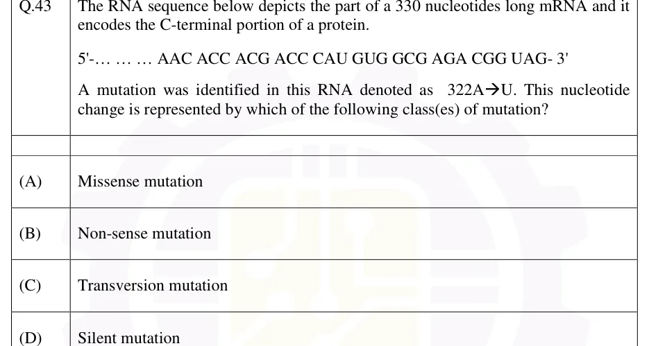
The RNA sequence below depicts the part of a 330 nucleotides long mRNA and it encodes the C-terminal portion of a protein.

5'-… … … AAC ACC ACG ACC CAU GUG GCG AGA CGG UAG-3'

A mutation was identified in this RNA denoted as 322A→U. This nucleotide change is represented by which of the following class(es) of mutation?

  1. (A)

    Missense mutation

  2. (B)

    Non-sense mutation

  3. (C)

    Transversion mutation

  4. (D)

    Silent mutation

Answer and solution
See the question in the paper
GATE 2026 BT Q.43: answer and solution

GATE 2025 BT Q.40

GATE 2025 · BTQ.40 · 2 marks · MCQ

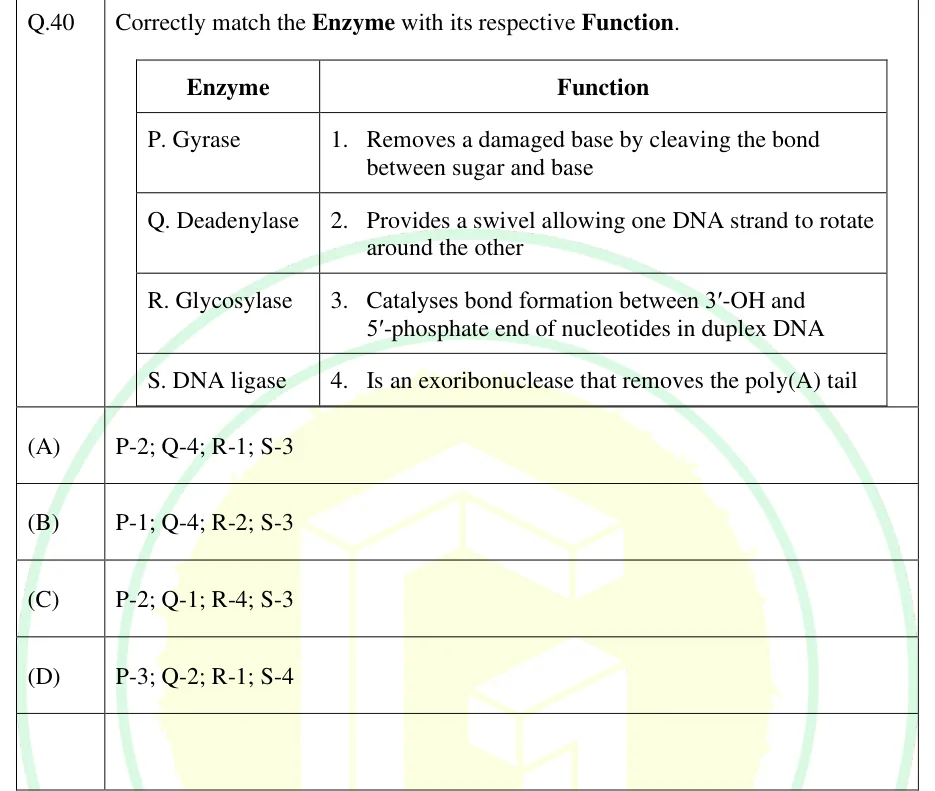
Correctly match the Enzyme with its respective Function.

Enzyme | Function
P. Gyrase | 1. Removes a damaged base by cleaving the bond between sugar and base
Q. Deadenylase | 2. Provides a swivel allowing one DNA strand to rotate around the other
R. Glycosylase | 3. Catalyses bond formation between 3'-OH and 5'-phosphate end of nucleotides in duplex DNA
S. DNA ligase | 4. Is an exoribonuclease that removes the poly(A) tail

GATE 2025 BT question 40: A two-column table titled 'Enzyme' and 'Function'.Tap to open full size
Figure from the GATE 2025 BT paper. It is part of the question.
Figure description

A two-column table titled 'Enzyme' and 'Function'. Four rows pair a lettered enzyme (P, Q, R, S) with a numbered function description (1, 2, 3, 4) as given in the stem above; no diagram, gel, pedigree or plot is present — it is purely a text-matching table.

  1. (A)

    P-2; Q-4; R-1; S-3

  2. (B)

    P-1; Q-4; R-2; S-3

  3. (C)

    P-2; Q-1; R-4; S-3

  4. (D)

    P-3; Q-2; R-1; S-4

Answer and solution
GATE 2025 BT Q.40: answer and solution

GATE 2023 BT Q.15

GATE 2023 · BTQ.15 · 1 mark · MCQ

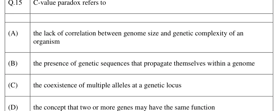
C-value paradox refers to

  1. (A)

    the lack of correlation between genome size and genetic complexity of an organism

  2. (B)

    the presence of genetic sequences that propagate themselves within a genome

  3. (C)

    the coexistence of multiple alleles at a genetic locus

  4. (D)

    the concept that two or more genes may have the same function

Answer and solution
See the question in the paper
GATE 2023 BT Q.15: answer and solution

GATE 2022 BT Q.46

GATE 2022 · BTQ.46 · 2 marks · MCQ

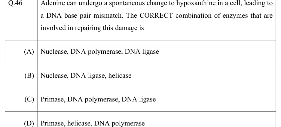
Adenine can undergo a spontaneous change to hypoxanthine in a cell, leading to a DNA base pair mismatch. The CORRECT combination of enzymes that are involved in repairing this damage is

  1. (A)

    Nuclease, DNA polymerase, DNA ligase

  2. (B)

    Nuclease, DNA ligase, helicase

  3. (C)

    Primase, DNA polymerase, DNA ligase

  4. (D)

    Primase, helicase, DNA polymerase

Answer and solution
See the question in the paper
GATE 2022 BT Q.46: answer and solution

PRACTICE

6 questions to test yourself

PRACTICE 11 mark · MCQ

In human cells, which DNA repair pathway removes UV-induced cyclobutane pyrimidine dimers and other bulky, helix-distorting lesions?

  1. (A)

    Base excision repair

  2. (B)

    Nucleotide excision repair

  3. (C)

    Mismatch repair

  4. (D)

    Direct reversal

Answer and solution

Answer: (B)

  1. A cyclobutane pyrimidine dimer covalently links adjacent pyrimidines on one strand and distorts the double helix. It is a bulky lesion, not a small chemical change to a single base.

  2. Direct reversal of pyrimidine dimers needs photolyase, which is absent in placental mammals, including humans. D cannot be the human pathway, although many bacteria do use it.

  3. Base excision repair uses DNA glycosylases that remove small, non-distorting base lesions (e.g., uracil, 8-oxoguanine). It does not remove dimers, so A is wrong.

  4. Mismatch repair corrects mismatches and small loops left by replication errors, not chemical damage, so C is wrong.

  5. Nucleotide excision repair recognises the helix distortion itself. It cuts the damaged strand on both sides, removes an oligonucleotide of about 24–32 nt in humans containing the lesion, and resynthesises it. Answer: B.

PRACTICE 21 mark · MCQ

A coding sequence has an insertion of 4 nucleotides within an exon, away from any splice site. What is the expected consequence?

  1. (A)

    Silent mutation

  2. (B)

    In-frame insertion, one extra amino acid added

  3. (C)

    Frameshift mutation, altering all downstream codons

  4. (D)

    No effect, since it lies within an exon

Answer and solution

Answer: (C)

  1. The mRNA is read in consecutive, non-overlapping triplets (codons), 5' to 3'.

  2. An insertion of a number of nucleotides that is not a multiple of 3 shifts the reading frame of every codon downstream of the insertion. This is a frameshift mutation.

  3. Every amino acid after the insertion site is changed, and a premature stop codon very often appears in the new frame.

  4. An insertion of exactly 3 (or a multiple of 3) nucleotides would add whole codons without shifting the frame, although it could still create a stop codon at the junction.

  5. 4 is not a multiple of 3, so the result is a frameshift. Answer: C.

PRACTICE 32 marks · MSQ

Which of the following statements about the Ames test are correct?

  1. (A)

    It scores reversion of histidine-auxotrophic Salmonella from his⁻ to his⁺

  2. (B)

    Rat liver (S9) extract is added to mimic mammalian metabolic activation of pro-mutagens

  3. (C)

    A reproducible, dose-dependent increase in revertant colonies above the spontaneous background indicates that the compound is mutagenic

  4. (D)

    The test directly measures tumour formation in live animals

Answer and solution

Answer: (A), (B), (C)

  1. The tester strains are histidine auxotrophs (his⁻) of Salmonella enterica serovar Typhimurium and cannot grow without histidine. They are plated on minimal medium with only a trace of histidine, enough for a few divisions but not for visible colonies. Cells whose his mutation reverts (his⁺) go on to form colonies, so A is correct.

  2. Many chemicals become mutagenic only after metabolism in the liver. The test is therefore run with and without rat liver S9 fraction (the 9000 g supernatant of liver homogenate, containing enzymes such as cytochrome P450) to supply this activation. B is correct.

  3. A reproducible, dose-dependent increase in revertant colonies over the spontaneous (control) count indicates that the compound is mutagenic in the test. C is correct.

  4. The test measures reversion mutations in bacteria. It does not measure tumour formation in animals; mutagenicity is only used as a screen for possible carcinogens. D is false.

  5. Answer: A, B and C.

PRACTICE 41 mark · NAT

A nucleosome core particle consists of a histone octamer with double-stranded DNA wrapped around it in about 1.65 left-handed superhelical turns. About how many base pairs of DNA are wrapped around the octamer in one core particle? (Give a whole number.)

Answer and solution

Answer: (accepted range )

  1. The core particle is the histone octamer (two copies each of H2A, H2B, H3 and H4) plus the DNA wrapped directly around it.

  2. High-resolution structures show about 147 bp of DNA in about 1.65 left-handed turns; the older, widely quoted value is 146 bp.

  3. Linker DNA between adjacent core particles, where histone H1 binds, is not part of the core-particle DNA.

  4. Answer: about 147 bp (146–148 accepted).

PRACTICE 51 mark · MCQ

A guanine in a G·C base pair is oxidised to 8-oxoguanine and is not repaired before replication. After two rounds of replication, which base-pair change is most commonly found in one of the descendant DNA molecules?

  1. (A)

    G·C → A·T transition

  2. (B)

    G·C → T·A transversion

  3. (C)

    A·T → G·C transition

  4. (D)

    A single-base-pair frameshift

Answer and solution

Answer: (B)

  1. 8-oxoguanine frequently pairs with A instead of C during replication.

  2. Round 1: the strand carrying 8-oxoG is copied and A is inserted opposite it, giving an 8-oxoG·A pair.

  3. Round 2: the new strand containing that A is copied and T is inserted opposite A, giving a T·A pair.

  4. The original G·C has become T·A. A purine has been replaced by a pyrimidine on the same strand (G to T), so this is a transversion.

  5. Answer: B.

PRACTICE 62 marks · MSQ

Which of the following statements about the repair of DNA double-strand breaks (DSBs) in eukaryotes are correct?

  1. (A)

    Homologous recombination (HR) is essentially error-free and uses a homologous template, usually the sister chromatid, which restricts it mainly to S/G2 phase

  2. (B)

    Non-homologous end joining (NHEJ) is error-prone and can act in G1 phase

  3. (C)

    Homologous recombination is the dominant double-strand break repair pathway in G1 phase

  4. (D)

    NHEJ requires extensive sequence homology between the two broken ends

Answer and solution

Answer: (A), (B)

  1. Homologous recombination copies the missing information from an intact homologous duplex, usually the sister chromatid. This makes it essentially error-free, but a sister chromatid exists only after replication, so HR works mainly in S and G2. A is correct.

  2. Non-homologous end joining ligates the broken ends directly, without a template. It can act in any phase, including G1, and often adds or loses a few nucleotides at the junction, so it is error-prone. B is correct.

  3. In G1 there is no sister chromatid, and NHEJ, not HR, is the predominant DSB repair pathway. C is false.

  4. NHEJ needs little or no sequence homology between the ends; extensive homology is a feature of HR. D is false.

  5. Answer: A and B.

OFFICIAL GATE 2027 SYLLABUS WORDING

Copied from the GATE 2027 syllabus; see all 2027 syllabi.

GATE BT

Molecular structure of genes and chromosomes; Mutations and mutagenesis … DNA damage and repair

Preparing in Kolkata? IAE Academy's course for GATE BT.