GATE 2026 BT Q.29
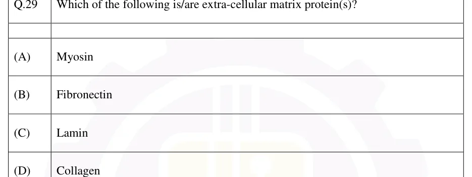
Which of the following is/are extra-cellular matrix protein(s)?
- (A)
Myosin
- (B)
Fibronectin
- (C)
Lamin
- (D)
Collagen

GATE BT
Cell structure, cell cycle, protein trafficking and cell death
Study notes for GATE BT "Cell structure, cell cycle, protein trafficking and cell death". Six sections cover eukaryotic organelles and the cytoskeleton; the cell cycle, CDK control, checkpoints and growth formulas; mitosis and meiosis with DNA-content bookkeeping; protein trafficking with sorting signals, glycosylation and vesicle coats; apoptosis, necrosis and autophagy; and the ECM, adhesion molecules and cell junctions. There are 6 practice questions (2 MCQ, 2 MSQ, 2 NAT), each answer recomputed independently.
15 previous-year questions · 15 solved · 6 practice questions
A eukaryotic cell is divided into membrane-bound compartments. Each compartment has its own set of enzymes, its own internal environment (for example, the acidic lysosome lumen) and its own function. Most proteins are encoded in the nucleus and made on cytosolic ribosomes. Sorting signals, covered in the protein trafficking section, send each protein to its compartment.
Major organelles and their principal functions
Organelle | Membrane | Principal function(s) |
|---|---|---|
Nucleus | Double (nuclear envelope, continuous with the ER; perforated by nuclear pore complexes; lined inside by the lamin-based nuclear lamina) | Genome storage, DNA replication, transcription and RNA processing. The nucleolus transcribes and processes rRNA and assembles the ribosomal subunits. |
Ribosome (cytosolic) | None | Protein synthesis. The eukaryotic cytosolic ribosome is 80S (60S + 40S); the bacterial ribosome is 70S (50S + 30S). Free ribosomes make cytosolic, nuclear, mitochondrial and peroxisomal proteins. ER-bound ribosomes make proteins for the secretory pathway. |
Rough ER | Single | Co-translational synthesis of proteins destined for secretion, the plasma membrane, the ER, the Golgi and lysosomes. N-linked glycosylation begins here, disulfide bonds form, and misfolded proteins are held back by quality control. |
Smooth ER | Single | Lipid and steroid synthesis, drug detoxification (cytochrome P450 enzymes, especially in liver), storage (the sarcoplasmic reticulum in muscle) |
Golgi apparatus | Single; stacked cisternae with a cis (entry) face and a trans (exit) face | Processing of N-linked oligosaccharides, O-linked glycosylation, sulfation, and mannose-6-phosphate tagging of lysosomal enzymes (in the cis-Golgi). Proteins are sorted and packaged at the trans-Golgi network (TGN). |
Lysosome | Single | Acid hydrolases working at a lumen pH of about 4.5–5, kept acidic by a V-type -ATPase. Lysosomes degrade material delivered by endocytosis, phagocytosis or autophagy. |
Peroxisome | Single; no DNA | Oxidases that generate , and catalase that breaks it down. In animal cells, peroxisomes carry out beta-oxidation of very-long-chain fatty acids, while most other fatty acids are oxidised in mitochondria. In plants and yeast, fatty-acid beta-oxidation is peroxisomal. Peroxisomes also make plasmalogens. Glyoxysomes are specialised plant peroxisomes that run the glyoxylate cycle. |
Mitochondrion | Double; inner membrane folded into cristae | Electron transport chain and ATP synthase in the inner membrane. Most TCA-cycle enzymes are in the matrix; succinate dehydrogenase (Complex II) sits in the inner membrane. The matrix also carries out fatty-acid beta-oxidation. Mitochondria have their own genome (circular mtDNA in animals) and bacteria-like ribosomes (55S in mammals), but most mitochondrial proteins are nuclear-encoded and imported. |
Chloroplast (plants, algae) | Double envelope, plus internal thylakoid membranes | Photosynthesis: the light reactions on the thylakoid membranes and carbon fixation (Calvin cycle) in the stroma. Chloroplasts have their own circular DNA and 70S ribosomes. |
Vacuole (plants) | Single (tonoplast) | Turgor pressure, storage of ions and metabolites, and lytic (lysosome-like) degradation |
The three cytoskeletal filament systems
Filament | Subunit | Approx. diameter | Polarity / nucleotide | Motor proteins | Standard drugs |
|---|---|---|---|---|---|
Microfilament (actin) | G-actin | about 7 nm | Polar (plus or barbed end grows faster than the minus or pointed end); binds ATP | Myosins | Cytochalasin blocks polymerisation; phalloidin stabilises filaments |
Microtubule | αβ-tubulin heterodimers; 13 protofilaments forming a hollow tube | about 25 nm | Polar; binds GTP; shows dynamic instability; nucleated at the MTOC (centrosome, γ-tubulin ring) | Kinesins (mostly plus-end directed), dyneins (minus-end directed) | Colchicine and nocodazole depolymerise; taxol (paclitaxel) stabilises |
Intermediate filament | Tissue-specific fibrous proteins: keratins, vimentin, neurofilament proteins, nuclear lamins | about 10 nm | Non-polar; no nucleotide binding | None | — |
Membrane structure: key points
Membranes follow the fluid mosaic model: a phospholipid bilayer with integral and peripheral proteins that can diffuse sideways within it.
Fluidity rises with more unsaturated (cis double-bonded) fatty acids and with shorter chains. Cholesterol lowers fluidity at higher temperatures and prevents tight packing (keeping the membrane fluid) at lower temperatures, so it buffers fluidity.
Lateral diffusion is fast. Transverse movement from one leaflet to the other ('flip-flop') is very slow unless catalysed by flippases, floppases or scramblases.
The membrane is asymmetric. In the plasma membrane, phosphatidylserine sits in the inner (cytosolic) leaflet and glycolipids and glycoproteins face outward. Phosphatidylserine appearing on the outer leaflet is an 'eat-me' signal in apoptosis.
Other structures to know
Cilia and flagella (eukaryotic) have a 9+2 microtubule axoneme (nine outer doublets around a central pair) and are driven by axonemal dynein. Their basal body, like a centriole, has nine triplets and no central pair.
Features of plant cells that animal cells lack: a cellulose cell wall, plastids, a large central vacuole and plasmodesmata. Higher plants also lack centrioles, yet still form a mitotic spindle.
Features of animal cells that plant cells lack: centrioles within the centrosome, and lysosomes in the classical sense (the plant vacuole performs the lytic role).
EXAM TIP
Common traps: catalase is a peroxisomal enzyme, not a lysosomal one. In animal cells most fatty-acid beta-oxidation is mitochondrial, and peroxisomes shorten very-long-chain fatty acids. Mitochondria and chloroplasts have double membranes and their own DNA; lysosomes and peroxisomes have single membranes and no DNA.
The cell cycle has four phases: G1, S (DNA synthesis), G2 and M (mitosis followed by cytokinesis). In a diploid cell the DNA content goes from 2C in G1 to 4C after S phase, and returns to 2C in each daughter cell. Cells that stop dividing leave G1 for a non-cycling state called G0. Cyclin-dependent kinases (CDKs) drive the cycle forward. CDK levels stay roughly constant, but a CDK is active only when bound to its cyclin, and cyclin levels rise and fall through the cycle. Phosphorylation and inhibitor proteins further tune CDK activity. Checkpoints stop the cycle until each step is complete and undamaged.
Cyclin–CDK combinations and their cell cycle role (mammalian cells)
Cyclin–CDK pair | Active during | Role |
|---|---|---|
Cyclin D–CDK4/6 | G1 | Cyclin D is made in response to mitogens (growth factors). The complex begins phosphorylating Rb, partly releasing E2F. |
Cyclin E–CDK2 | Late G1 to the G1/S transition | Completes Rb hyperphosphorylation. Because E2F induces cyclin E, this is a positive-feedback loop. The complex commits the cell to S phase and triggers replication origin firing. |
Cyclin A–CDK2 | S phase | Drives DNA replication and helps prevent re-replication. Cyclin A also pairs with CDK1 in G2. |
Cyclin B–CDK1 | G2/M | Drives entry into mitosis: chromosome condensation, nuclear envelope breakdown and spindle assembly. The complex is also called MPF (maturation- or M-phase-promoting factor). |
How CDK activity is controlled
Cyclin binding is required for any activity.
An activating phosphorylation (on Thr161 of CDK1) is added by CDK-activating kinase (CAK).
An inhibitory phosphorylation (on Tyr15 of CDK1) is added by Wee1 kinase and removed by Cdc25 phosphatase. Removal by Cdc25 is what triggers the burst of cyclin B–CDK1 activity at mitotic entry.
CDK inhibitor proteins (CKIs) block activity: INK4 proteins such as p16 inhibit CDK4/6, and Cip/Kip proteins such as p21 and p27 inhibit cyclin E–CDK2 and cyclin A–CDK2. The p53 protein induces p21.
Cyclins are destroyed by ubiquitin-mediated proteolysis: SCF targets G1/S cyclins and CKIs such as p27, and APC/C targets securin and the mitotic cyclins (A and B).
Cell cycle checkpoints, in order
G1/S checkpoint (the restriction point in mammalian cells, called 'Start' in yeast). It checks cell size, nutrients and growth factors, and DNA integrity before the cell commits to replication. It is controlled by the Rb–E2F switch. DNA damage activates p53, which blocks passage through p21.
Intra-S checkpoint: replication stress or damage during S phase slows origin firing.
G2/M checkpoint: it checks that replication is complete and the DNA undamaged before mitosis. DNA damage activates ATM (double-strand breaks) or ATR (single-stranded DNA and replication stress), which activate Chk2 or Chk1. These inhibit Cdc25, so cyclin B–CDK1 stays in its inhibited, Tyr15-phosphorylated state.
Spindle assembly checkpoint, at the metaphase-to-anaphase transition: an unattached kinetochore generates a signal (involving Mad2) that inhibits APC/C–Cdc20. Once every kinetochore is attached, APC/C–Cdc20 ubiquitinates securin and cyclin B for proteasomal degradation. Separase is then freed to cleave cohesin, the sister chromatids separate, and the fall in CDK1 activity allows exit from mitosis.
EXPONENTIAL GROWTH OF A CELL POPULATION
Here is the initial cell count, the count after time , and the doubling time. It holds only while every cell is cycling with the same doubling time, with no cell death and outside any lag or stationary phase.
NUMBER OF GENERATIONS (DOUBLINGS) IN TIME T
This follows directly from the exponential-growth formula under the same conditions.
SPECIFIC GROWTH RATE
This is the same exponential law written with base . has units of reciprocal time.
PHASE DURATION FROM THE FRACTION OF CELLS IN THAT PHASE (E.G., MITOTIC INDEX)
is the total cell-cycle time and is the fraction of cells observed in the phase; for M phase this fraction is the mitotic index. This is an approximation for an asynchronous culture that treats cells as evenly spread across cycle ages. In an exponentially growing culture young cells slightly outnumber old ones, so the estimate is not exact.
WORKED EXAMPLE
In an asynchronously growing cell culture with a cell-cycle time of 25 h, 40 of 1,000 cells counted are in mitosis. Estimate the duration of M phase.
Mitotic index = 40/1000 = 0.04.
h.
Answer: About 1 h
Two classes of genes control cell growth. Tumour suppressors act as brakes. p53 responds to DNA damage by arresting the cycle, at G1/S through p21 and also at G2/M, or by triggering apoptosis if the damage cannot be repaired. Normally the ubiquitin ligase MDM2 keeps p53 levels low; MDM2 is itself induced by p53, forming a negative-feedback loop. Rb binds E2F and blocks the G1/S transition. Proto-oncogenes, such as Ras, Myc and cyclin D, act as accelerators. At the level of the cell, tumour suppressors behave recessively, because both alleles must be lost ('two-hit'), while oncogenes behave dominantly, because one gain-of-function allele is enough. A proto-oncogene becomes an oncogene through gain-of-function mutation, amplification or translocation.
EXAM TIP
Common traps: losing a tumour suppressor such as p53 or Rb, not gaining one, drives uncontrolled division, and the logic is the reverse for oncogenes. Also, CDK protein levels stay fairly constant; it is the cyclin levels that oscillate.
Mitosis produces two daughter cells that are genetically identical to the parent and have the same chromosome number, for growth and repair. Meiosis is two rounds of division (meiosis I and II) after a single round of DNA replication. It halves the chromosome number of a diploid cell, producing four haploid cells that differ genetically. The variation comes from two mechanisms mitosis lacks: crossing over between homologous chromosomes and independent assortment of homologues.
Stages of mitosis, in order
Prophase: chromatin condenses into chromosomes made of two sister chromatids, the centrosomes separate and the spindle begins to form, and the nucleolus disappears.
Prometaphase: the nuclear envelope breaks down (in animal cells; some fungi have a 'closed' mitosis) and spindle microtubules attach to the kinetochores.
Metaphase: the chromosomes line up at the metaphase plate.
Anaphase: cohesin is cleaved and sister chromatids separate. In anaphase A they move to the poles as kinetochore microtubules shorten; in anaphase B the poles move apart.
Telophase: the nuclear envelopes re-form and the chromosomes decondense.
Cytokinesis: in animal cells a contractile ring of actin and myosin II forms a cleavage furrow. In plant cells a phragmoplast guides Golgi-derived vesicles to build a cell plate.
Mitosis vs meiosis (meiosis shown for a diploid, 2n, parent)
Feature | Mitosis | Meiosis |
|---|---|---|
Rounds of DNA replication / divisions | One / one | One / two (meiosis I, meiosis II); no replication between them |
Homologue pairing (synapsis) | Normally absent | Present in prophase I (synaptonemal complex) |
Crossing over | Normally absent (mitotic recombination is rare) | Present in prophase I, between non-sister chromatids |
Homologue separation | Does not occur | Anaphase I (the reductional division) |
Sister chromatid separation | Anaphase | Anaphase II (the equational division). Cohesin at the centromeres is protected in meiosis I until anaphase II. |
Products | 2 cells with the parent's chromosome number, genetically identical to it | 4 haploid (n) cells, genetically different from each other and from the parent |
Chromosome number and DNA content through meiosis (diploid 2n; C = haploid DNA content)
Stage | Chromosomes per cell | Chromatids per chromosome | DNA content per cell |
|---|---|---|---|
G1 (before replication) | 2n | 1 | 2C |
After S phase (G2, prophase I) | 2n | 2 | 4C |
Each cell after meiosis I | n | 2 | 2C |
Each cell after meiosis II | n | 1 | 1C |
Each daughter cell of mitosis (for comparison) | 2n | 1 (after division) | 2C |
Meiosis I prophase substages, in order
Leptotene: chromosomes begin to condense.
Zygotene: homologous chromosomes begin pairing (synapsis) and the synaptonemal complex forms.
Pachytene: synapsis is complete and crossing over takes place between non-sister chromatids.
Diplotene: the synaptonemal complex dissolves and homologues stay joined at chiasmata, which become visible. Oocytes of many animals, including humans, arrest for a long period at this stage (the dictyotene stage).
Diakinesis: chromosomes condense further, the chiasmata move toward the chromosome ends, and the nuclear envelope breaks down in preparation for metaphase I.
INDEPENDENT ASSORTMENT
Here is the haploid chromosome number. The count ignores crossing over, which adds further variation. For humans (), .
Nondisjunction
Nondisjunction in meiosis I, when a pair of homologues fails to separate, makes all four gametes abnormal: two are n + 1 and two are n − 1 for that chromosome.
Nondisjunction in meiosis II, when sister chromatids fail to separate in one of the two cells, gives two normal gametes, one n + 1 and one n − 1.
Fertilisation of an n + 1 gamete by a normal gamete gives a trisomic zygote (2n + 1); an n − 1 gamete gives a monosomic zygote (2n − 1).
EXAM TIP
Common traps: meiosis I is the reductional division because homologues separate, halving the chromosome number, while meiosis II is equational. After meiosis I each cell is already haploid (n) but still holds 2C DNA, because each chromosome still has two chromatids.
A protein's destination is encoded in sorting signals within its sequence, or in modifications added to it. All proteins start being made on cytosolic ribosomes, and then follow one of two targeting routes. (1) Proteins bound for the ER, Golgi, lysosomes, plasma membrane or secretion are usually sent to the ER co-translationally by a signal sequence. Post-translational import into the ER also exists, especially in yeast. (2) Proteins for the nucleus, mitochondria, chloroplasts and peroxisomes are finished on free ribosomes and imported post-translationally through dedicated pores or translocons, not in vesicles. Secretory-pathway proteins travel in vesicles along the route ER → ER–Golgi intermediate compartment (ERGIC) → cis-Golgi → medial-Golgi → trans-Golgi → trans-Golgi network → final destination. Retrograde COPI transport returns escaped ER proteins.
Secretory (co-translational) pathway, in order
The signal recognition particle (SRP) recognises a hydrophobic N-terminal signal sequence on the growing chain, pauses elongation, and docks the ribosome on the SRP receptor at the rough ER. This docking is GTP-dependent.
The chain enters the ER lumen through the Sec61 translocon, and signal peptidase usually cleaves the signal sequence. In membrane proteins, stop-transfer and internal signal-anchor sequences set the orientation and number of membrane crossings. Some proteins receive a GPI anchor in the ER lumen.
Correctly folded cargo leaves the ER in COPII-coated vesicles for the ERGIC and cis-Golgi.
Proteins are modified in sequence through the Golgi from cis to trans. ER-resident proteins that escape are retrieved in COPI-coated vesicles: soluble proteins by a C-terminal KDEL signal bound by the KDEL receptor, and membrane proteins by a C-terminal cytosolic KKXX (dilysine) signal bound directly by COPI.
In the cis-Golgi, lysosomal hydrolases are tagged with mannose-6-phosphate (M6P). In the trans-Golgi network, M6P receptors package them into clathrin-coated vesicles bound for late endosomes. There the acidic pH releases the enzymes, which reach lysosomes, and the receptors recycle. Failure of M6P tagging (I-cell disease) causes lysosomal enzymes to be secreted instead.
Other proteins leave the trans-Golgi network by constitutive secretion, the default route that needs no signal, or by regulated secretion from secretory granules in response to a signal.
Glycosylation and ER quality control
N-linked glycosylation: a pre-assembled oligosaccharide () on dolichol phosphate is transferred in one piece by oligosaccharyltransferase to Asn in the sequence Asn-X-Ser/Thr, where X is any amino acid except Pro. This happens in the ER lumen, usually while the chain is still being made. The oligosaccharide is then trimmed and remodelled in the ER and Golgi.
O-linked glycosylation: sugars are attached to the hydroxyl of Ser or Thr, mainly in the Golgi.
Quality control: the lectin chaperones calnexin and calreticulin hold glycoproteins in the ER until they fold. Proteins that stay misfolded are moved back into the cytosol, ubiquitinated and degraded by the proteasome (ER-associated degradation, ERAD). Accumulated unfolded protein triggers the unfolded protein response (UPR).
Sorting signals and their destinations
Signal/tag | Destination | Machinery |
|---|---|---|
N-terminal hydrophobic ER signal sequence | Co-translational entry into the ER | SRP, SRP receptor, Sec61 translocon (not a vesicle) |
C-terminal KDEL (HDEL in yeast), on soluble proteins | Retrieval from the Golgi or ERGIC back to the ER | KDEL receptor, COPI vesicles (retrograde) |
C-terminal KKXX, on the cytosolic tail of membrane proteins | Retrieval to the ER | Bound directly by COPI (retrograde) |
Mannose-6-phosphate (a carbohydrate tag, not a peptide) | Trans-Golgi network → late endosome → lysosome | M6P receptor, clathrin-coated vesicles |
Nuclear localisation signal (NLS; short internal stretch rich in basic Lys/Arg residues) | Cytosol to nucleus, through the nuclear pore complex; proteins are imported folded | Importins; a Ran-GTP (nucleus) / Ran-GDP (cytosol) gradient releases the cargo in the nucleus (not vesicular) |
Nuclear export signal (NES) | Nucleus to cytosol | Exportins, Ran-GTP |
N-terminal mitochondrial presequence (positively charged amphipathic α-helix) | Cytosol to the mitochondrial matrix, post-translationally | TOM (outer membrane) and TIM23 (inner membrane) complexes. The protein is imported unfolded, which requires the inner-membrane potential and ATP (matrix Hsp70). The presequence is then cleaved by the matrix processing peptidase. |
N-terminal chloroplast transit peptide | Cytosol to the chloroplast stroma, post-translationally | TOC and TIC complexes |
C-terminal Ser-Lys-Leu (SKL; PTS1), or N-terminal PTS2 | Cytosol to the peroxisome matrix; proteins are imported folded | Peroxin (Pex) import receptors |
Vesicle coats
Coat | Recruiting GTPase / adaptors | Route |
|---|---|---|
COPII | Sar1 | ER → ERGIC / cis-Golgi (anterograde) |
COPI | ARF1 | Golgi → ER (retrograde) and between Golgi cisternae |
Clathrin | ARF1 with the AP1 adaptor at the TGN; AP2 at the plasma membrane; dynamin pinches the vesicle off | TGN → endosomes; plasma membrane → endosomes (receptor-mediated endocytosis) |
Rab GTPases and tethering proteins ensure a vesicle docks at the right membrane. Pairing of a v-SNARE on the vesicle with t-SNAREs on the target membrane drives fusion, and NSF with SNAPs then separates the SNARE complex so the SNAREs can be reused.
EXAM TIP
Import into the nucleus, mitochondria, chloroplasts and peroxisomes is post-translational and uses no COPI, COPII or clathrin vesicles; each organelle has its own pore or translocon. Two further contrasts: mitochondria import proteins unfolded, while nuclear pores and peroxisomes import folded proteins. KDEL retrieval uses COPI (retrograde), not COPII.
Apoptosis is programmed, energy-dependent cell death carried out by a caspase cascade. Phagocytes remove the dying cell without inflammation. Accidental necrosis is uncontrolled death after acute injury: the cell swells and its membrane ruptures, causing inflammation. Some forms of necrotic death are regulated, however. Necroptosis runs through RIPK1, RIPK3 and MLKL, does not need caspases, and is favoured when caspase-8 is inhibited. Pyroptosis is inflammatory, depends on caspase-1 and makes membrane pores of gasdermin D. Autophagy is a regulated, lysosome-dependent recycling pathway that usually helps cells survive stress, but can contribute to cell death when prolonged.
Apoptosis vs necrosis vs autophagy
Feature | Apoptosis | Necrosis (accidental) | Autophagy |
|---|---|---|---|
Trigger | Developmental cues, DNA damage, growth-factor withdrawal, death-receptor signalling | Acute injury (ischaemia, toxins, trauma) | Nutrient starvation, damaged organelles, protein aggregates, stress |
ATP requirement | Required (active process) | Not required (ATP depletion is often the cause) | Required |
Cell volume | Shrinks | Swells | No characteristic change |
Key machinery | Caspases (initiator and executioner) | No dedicated machinery; ion-pump failure, membrane rupture | ATG proteins build the double-membrane autophagosome, which fuses with the lysosome |
Membrane | Stays intact until engulfment; blebbing, apoptotic bodies, phosphatidylserine exposed on the outer leaflet ('eat-me' signal) | Ruptures early (lysis) | Stays intact; cargo is enclosed in a double-membrane autophagosome |
Inflammation | Absent (the cell is engulfed before it breaks open) | Present (cell contents released) | Absent |
Chromatin / DNA | Condensation (pyknosis). Internucleosomal cleavage by the caspase-activated DNase (CAD) gives a 'ladder' of fragments in multiples of about 180–200 bp. | Random degradation (a smear on a gel) | No characteristic chromatin change |
Apoptosis: extrinsic and intrinsic pathways
Caspases are cysteine proteases that cut after an aspartate residue. They are made as inactive procaspases. Initiator caspases (-8, -9, and -10 in humans) are activated by being brought together on a scaffold. Executioner caspases (-3, -6, -7) are activated when initiators cleave them.
Extrinsic (death-receptor) pathway: a ligand such as FasL or TNF binds its death receptor (Fas, TNFR1). The adaptor FADD then assembles the death-inducing signalling complex (DISC), which activates caspase-8.
Intrinsic (mitochondrial) pathway: stress or DNA damage activates BH3-only proteins (e.g., Bid, Bim, Bad, Puma, Noxa). These overcome the anti-apoptotic proteins Bcl-2, Bcl-xL and Mcl-1 and activate the effectors Bax and Bak, which permeabilise the outer mitochondrial membrane.
Cytochrome c released into the cytosol binds Apaf-1, which, with dATP or ATP, assembles into the apoptosome. The apoptosome activates caspase-9. Smac/DIABLO, released from mitochondria at the same time, neutralises the inhibitor-of-apoptosis proteins (IAPs, e.g., XIAP).
The two pathways are linked: caspase-8 cleaves Bid to tBid, which activates Bax and Bak and amplifies the extrinsic signal through the mitochondria.
Both pathways converge on executioner caspases, mainly caspase-3. These cleave structural and nuclear proteins, and cleave ICAD to free CAD for DNA fragmentation, producing the apoptotic morphology.
Autophagy: key points
Types: macroautophagy (a double-membrane autophagosome encloses the cargo), microautophagy (the lysosome membrane takes up cargo directly) and chaperone-mediated autophagy (proteins with a KFERQ-like motif are recognised by Hsc70 and moved across the lysosome membrane via LAMP-2A).
Regulation: when nutrients are plentiful, mTORC1 suppresses autophagy by inhibiting the ULK1 complex. Starvation, or the drug rapamycin, inhibits mTORC1, and AMPK activation at low energy promotes autophagy.
The Beclin-1–VPS34 (class III PI3-kinase) complex starts the autophagosome membrane.
LC3 (Atg8) is converted to lipid-linked LC3-II, which binds the autophagosome membrane and is a standard marker of autophagosomes.
The autophagosome fuses with a lysosome to form an autolysosome, where the cargo is degraded and its building blocks recycled.
EXAM TIP
Common traps: it is necrosis that is inflammatory and apoptosis that is not. Apoptotic cells shrink while necrotic cells swell. Autophagy delivers the cell's own cytoplasmic material to the lysosome in an autophagosome; it is not phagocytosis of a whole cell. Caspase-9 belongs to the intrinsic pathway, caspase-8 to the extrinsic pathway, and caspase-3 is an executioner.
The extracellular matrix (ECM) is the network of proteins and polysaccharides outside cells. It gives structural support and also signals to cells. Cells attach to the ECM and to each other through adhesion molecules, which pass signals into the cell that affect survival, growth and division. Most normal cells, other than blood cells, show anchorage dependence: they must be attached to the ECM, mainly through integrin signalling, to proliferate, and detached cells often undergo a form of apoptosis called anoikis. Normal cells in culture also show contact inhibition, stopping division once they are confluent. Transformed (cancer) cells commonly lose both controls.
Major ECM components
Collagen is the most abundant protein in mammals. It is a triple helix of three chains built from Gly-X-Y repeats, in which every third residue is glycine, X is often proline and Y often 4-hydroxyproline. Prolyl hydroxylation needs ascorbate (vitamin C), whose deficiency causes scurvy. Collagen is secreted as procollagen, its propeptides are cut off outside the cell, and lysyl oxidase cross-links it. Fibrillar collagens (e.g., type I in skin, bone and tendon; type II in cartilage) give tensile strength. Type IV collagen forms a sheet-like network in the basal lamina instead of fibrils.
Elastin forms a cross-linked network (via desmosine and isodesmosine, made from lysine residues) that gives elastic recoil. It is laid down on fibrillin microfibrils.
Glycosaminoglycans (GAGs) and proteoglycans are negatively charged polysaccharides that bind water and resist compression. In proteoglycans, sulfated GAGs such as heparan sulfate and chondroitin sulfate are attached to a core protein. Hyaluronan is the exception: it is not sulfated, is not attached to a core protein, and is made at the plasma membrane rather than in the Golgi.
Fibronectin is a dimeric glycoprotein that links cells to the matrix. Its RGD (Arg-Gly-Asp) sequence binds integrins.
Laminin is a cross-shaped αβγ heterotrimer and a major component of the basal lamina. It links cells to the type IV collagen network.
In plants the extracellular matrix is the cell wall: cellulose microfibrils embedded in hemicellulose and pectin.
Cell adhesion molecule classes
Class | Binding partner | Cation dependence | Example role |
|---|---|---|---|
Integrins (α/β heterodimers) | ECM proteins (fibronectin via RGD, laminin, collagen). Some leukocyte integrins bind immunoglobulin-superfamily ligands such as ICAM-1. | Divalent-cation dependent | Cell–ECM adhesion. Integrins connect to actin through talin and vinculin at focal adhesions, and to intermediate filaments at hemidesmosomes. They signal in both directions across the membrane. |
Cadherins | The same cadherin on a neighbouring cell (homophilic) | -dependent | Cell–cell adhesion. Classical cadherins link to actin through catenins at adherens junctions; desmosomal cadherins (desmogleins, desmocollins) link to intermediate filaments at desmosomes. |
Selectins | Carbohydrate ligands on other cells (lectin binding) | -dependent | Brief cell–cell adhesion, e.g., leukocytes rolling on endothelium |
Immunoglobulin superfamily (e.g., NCAM, ICAM) | Homophilic or heterophilic protein–protein binding (e.g., ICAM-1 with an integrin) | -independent | Cell–cell adhesion in the nervous and immune systems |
Cell junctions
Junction | Main proteins | Cytoskeletal link | Function |
|---|---|---|---|
Tight junction | Claudins, occludin | Actin (via adaptors) | Seals the gap between cells (paracellular barrier) and keeps apical and basolateral membrane proteins apart |
Adherens junction | Classical cadherins, catenins | Actin | Cell–cell anchoring |
Desmosome | Desmosomal cadherins, plakoglobin, desmoplakin | Intermediate filaments (keratins in epithelia) | Strong cell–cell anchoring that resists mechanical stress |
Gap junction | Connexins (six form a connexon; connexons from two cells align) | None | Direct passage of ions and small molecules up to about 1 kDa between neighbouring cells |
Hemidesmosome | Integrin α6β4, binding laminin | Intermediate filaments | Cell–basal lamina anchoring |
Focal adhesion | Integrins, talin, vinculin | Actin | Cell–ECM anchoring and signalling |
Plasmodesma (plants) | Channel through the cell wall lined by plasma membrane, with ER-derived desmotubule | — | Cytoplasmic continuity between plant cells |
EXAM TIP
Common traps: cadherins and selectins are -dependent, while immunoglobulin-superfamily adhesion molecules are not. Desmosomes and hemidesmosomes anchor intermediate filaments, whereas adherens junctions and focal adhesions anchor actin. Hyaluronan is the one glycosaminoglycan that is neither sulfated nor bound to a core protein.
PREVIOUS-YEAR QUESTIONS
Try each question first, then open the answer. We placed each question under a topic ourselves; the exam paper does not label topics.
See these papers in full: GATE BT 2026 · GATE BT 2025 · GATE BT 2024 · GATE BT 2023 · GATE BT 2022 · GATE BT 2020 · GATE BT 2019
Which of the following is/are extra-cellular matrix protein(s)?
Myosin
Fibronectin
Lamin
Collagen
In the process of generating a clonal mammalian cell line, a single cell was seeded in a well of a cell culture plate. After the first 48 hours, one of the progeny cells underwent apoptosis due to a new mutation. If the doubling time of the cells is 24 hours and no more cell death occurs, the total number of cells after a total of 7 days from seeding will be ________. (answer in integer)
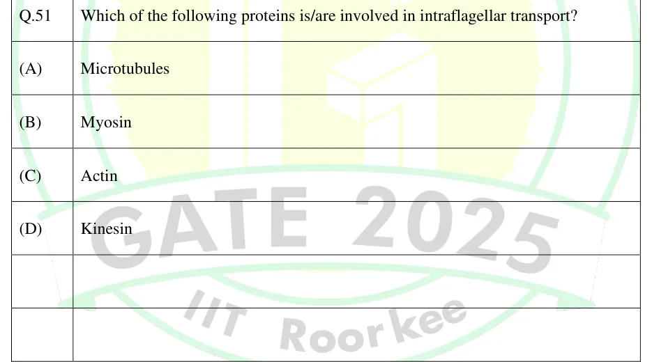
Which of the following proteins is/are involved in intraflagellar transport?
Microtubules
Myosin
Actin
Kinesin
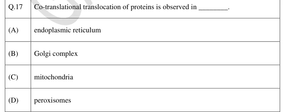
Co-translational translocation of proteins is observed in ________.
endoplasmic reticulum
Golgi complex
mitochondria
peroxisomes
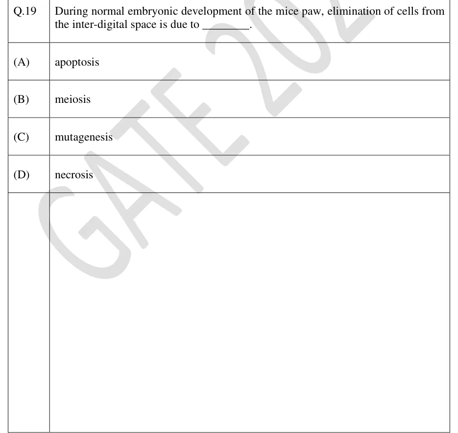
During normal embryonic development of the mice paw, elimination of cells from the inter-digital space is due to ________.
apoptosis
meiosis
mutagenesis
necrosis
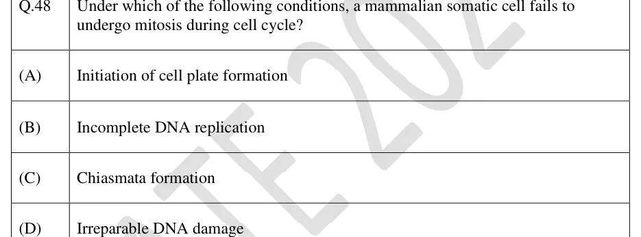
Under which of the following conditions, a mammalian somatic cell fails to undergo mitosis during cell cycle?
Initiation of cell plate formation
Incomplete DNA replication
Chiasmata formation
Irreparable DNA damage
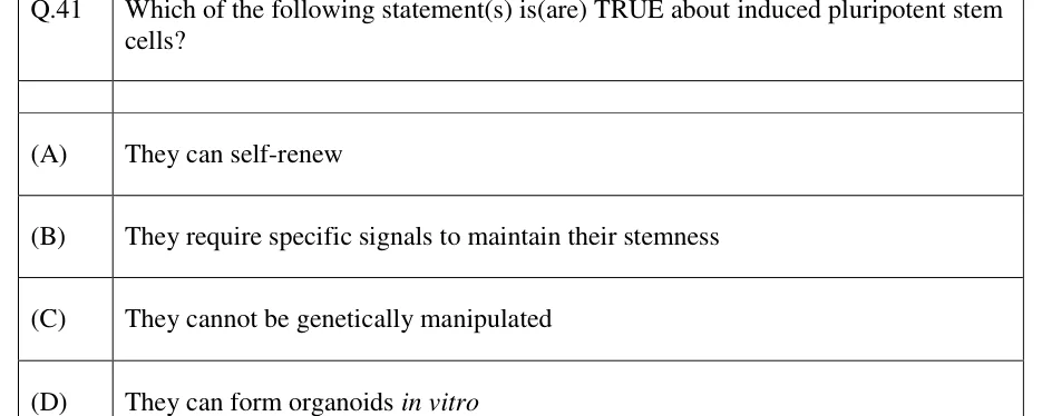
Which of the following statement(s) is(are) TRUE about induced pluripotent stem cells?
They can self-renew
They require specific signals to maintain their stemness
They cannot be genetically manipulated
They can form organoids in vitro
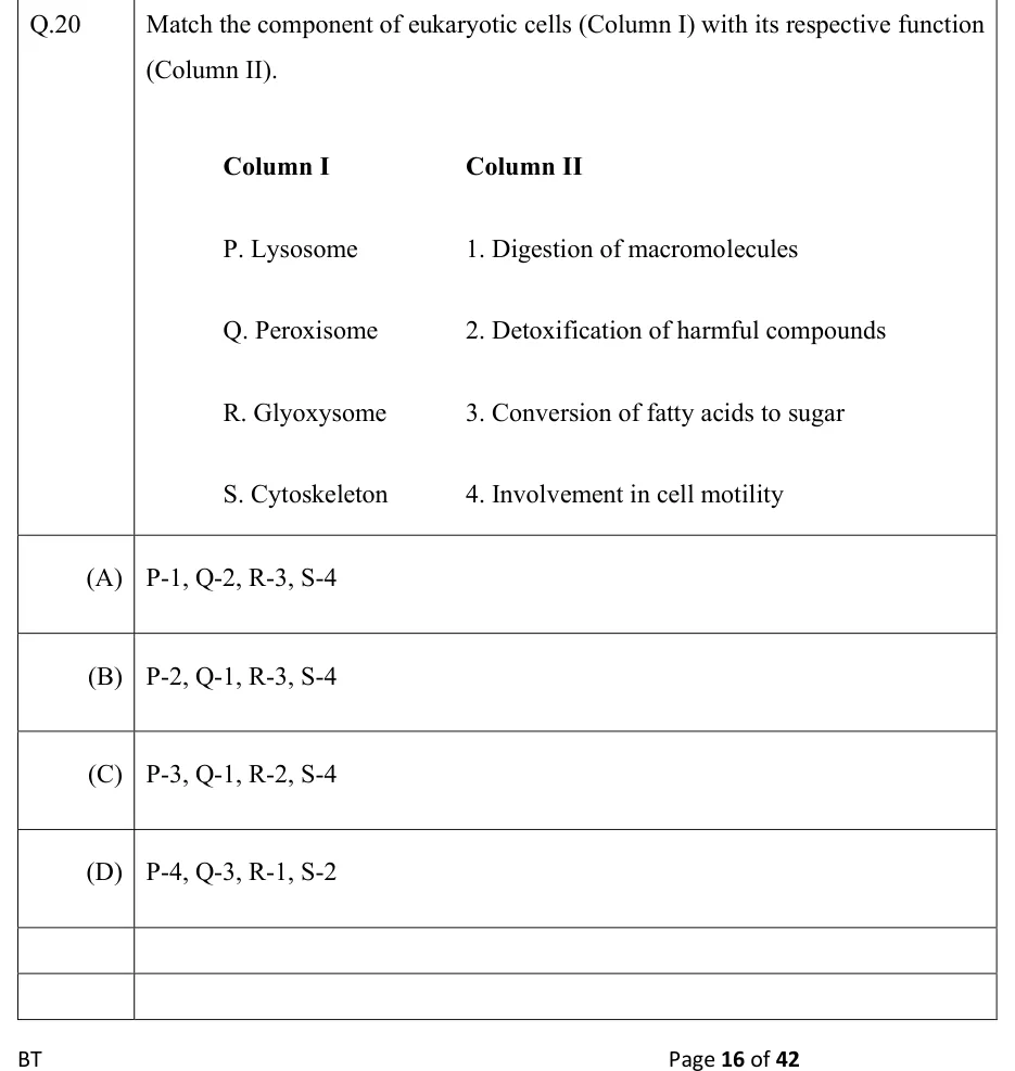
Match the component of eukaryotic cells (Column I) with its respective function (Column II).
Column I: P. Lysosome, Q. Peroxisome, R. Glyoxysome, S. Cytoskeleton
Column II: 1. Digestion of macromolecules, 2. Detoxification of harmful compounds, 3. Conversion of fatty acids to sugar, 4. Involvement in cell motility
P-1, Q-2, R-3, S-4
P-2, Q-1, R-3, S-4
P-3, Q-1, R-2, S-4
P-4, Q-3, R-1, S-2
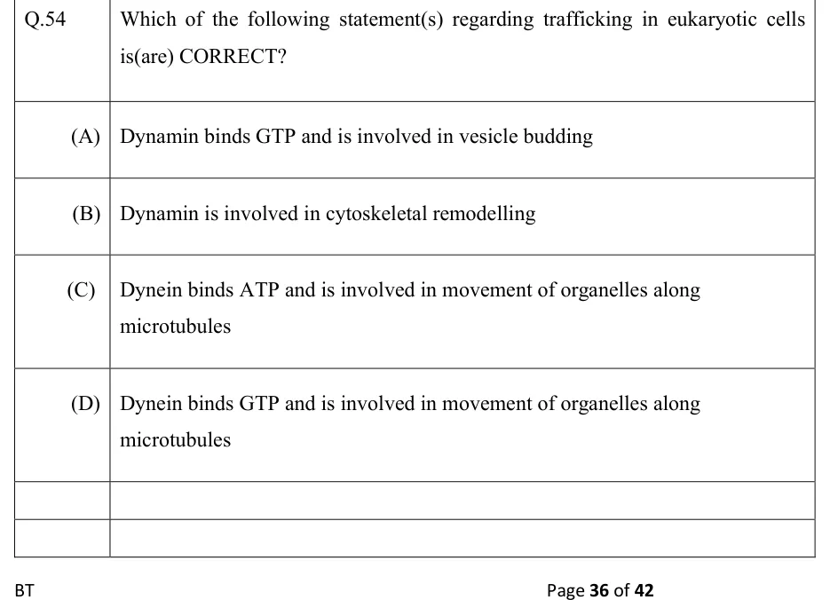
Which of the following statement(s) regarding trafficking in eukaryotic cells is(are) CORRECT?
Dynamin binds GTP and is involved in vesicle budding
Dynamin is involved in cytoskeletal remodelling
Dynein binds ATP and is involved in movement of organelles along microtubules
Dynein binds GTP and is involved in movement of organelles along microtubules
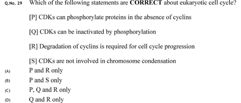
Which of the following statements are CORRECT about eukaryotic cell cycle? P CDKs can phosphorylate proteins in the absence of cyclins. Q CDKs can be inactivated by phosphorylation. R Degradation of cyclins is required for cell cycle progression. S CDKs are not involved in chromosome condensation.
P and R only
P and S only
P, Q and R only
Q and R only
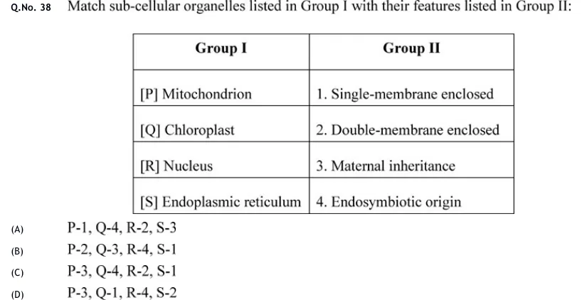
Match sub-cellular organelles listed in Group I with their features listed in Group II:
Group I: [P] Mitochondrion, [Q] Chloroplast, [R] Nucleus, [S] Endoplasmic reticulum
Group II: 1. Single-membrane enclosed, 2. Double-membrane enclosed, 3. Maternal inheritance, 4. Endosymbiotic origin
 Tap to open full size
A two-column table. Column header 'Group I' lists: [P] Mitochondrion, [Q] Chloroplast, [R] Nucleus, [S] Endoplasmic reticulum. Column header 'Group II' lists: 1. Single-membrane enclosed, 2. Double-membrane enclosed, 3. Maternal inheritance, 4. Endosymbiotic origin. Rows are aligned P-1, Q-2, R-3, S-4 by position but these are just the listed items, not the answer.
P-1, Q-4, R-2, S-3
P-2, Q-3, R-4, S-1
P-3, Q-4, R-2, S-1
P-3, Q-1, R-4, S-2
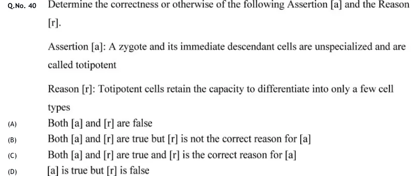
Determine the correctness or otherwise of the following Assertion [a] and the Reason [r].
Assertion [a]: A zygote and its immediate descendant cells are unspecialized and are called totipotent
Reason [r]: Totipotent cells retain the capacity to differentiate into only a few cell types
Both [a] and [r] are false
Both [a] and [r] are true but [r] is not the correct reason for [a]
Both [a] and [r] are true and [r] is the correct reason for [a]
[a] is true but [r] is false
Assume that a cell culture was started with five human fibroblast cells. Two cells did not divide even once whereas the other three cells completed three rounds of cell division. At this stage, the total number of kinetochores in all the cells put together is ______.
DNA synthesis in eukaryotes occurs during which phase of the mitotic cell cycle?
M
G_1
S
G_0
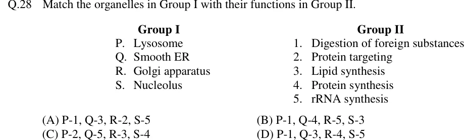
Match the organelles in Group I with their functions in Group II.
Group I:
P. Lysosome
Q. Smooth ER
R. Golgi apparatus
S. Nucleolus
Group II:
1. Digestion of foreign substances
2. Protein targeting
3. Lipid synthesis
4. Protein synthesis
5. rRNA synthesis
P-1, Q-3, R-2, S-5
P-1, Q-4, R-5, S-3
P-2, Q-5, R-3, S-4
P-1, Q-3, R-4, S-5
PRACTICE
In a eukaryotic cell, the electron transport chain and oxidative phosphorylation are located in which organelle?
Rough endoplasmic reticulum
Mitochondrion
Golgi apparatus
Peroxisome
Answer: (B)
Oxidative phosphorylation needs the electron transport chain complexes and ATP synthase.
Both are embedded in the inner mitochondrial membrane. The matrix contains most TCA-cycle enzymes, which supply NADH and to the chain; succinate dehydrogenase (Complex II) is itself in the inner membrane.
The rough ER, Golgi and peroxisome carry no electron transport chain complexes coupled to ATP synthesis.
Which of the following are recognised surveillance checkpoints of the eukaryotic cell cycle?
G1/S checkpoint
G2/M checkpoint
Spindle assembly checkpoint
G0 checkpoint
Answer: (A), (B), (C)
G1/S checkpoint: checks cell size, nutrients and growth factors, and DNA integrity before the cell commits to replication (Rb–E2F, p53–p21).
G2/M checkpoint: checks that DNA replication is complete and the DNA undamaged before mitosis (ATM/ATR → Chk1/Chk2 → inhibition of Cdc25).
Spindle assembly checkpoint (metaphase-to-anaphase): checks that every kinetochore is attached to the spindle before APC/C–Cdc20 allows sister chromatids to separate.
G0 is a quiescent, non-cycling state, not a checkpoint.
A mammalian cell line is growing exponentially in culture, with every cell cycling and no cell death. The population is cells at and cells at hours. Using , calculate the doubling time in hours.
Answer: (accepted range )
.
So , giving (three generations).
hours.
A soluble ER-resident protein that has escaped to the Golgi is returned to the ER. Which signal and transport step are responsible for this retrieval?
N-terminal signal sequence, co-translational insertion via SRP
C-terminal KDEL signal, retrograde COPI vesicle transport
C-terminal KDEL signal, anterograde COPII vesicle transport
Mannose-6-phosphate tag, clathrin-coated vesicle transport
Answer: (B)
Soluble ER-resident proteins carry a C-terminal KDEL signal (HDEL in yeast).
The KDEL receptor in the ERGIC and cis-Golgi binds this signal.
The receptor–cargo complex returns to the ER in COPI-coated vesicles (retrograde transport). COPII vesicles carry cargo the other way, from the ER to the Golgi. Clathrin vesicles work after the Golgi and in endocytosis, and M6P directs lysosomal enzymes, not ER retrieval.
Option A describes entry into the ER, not retrieval from the Golgi.
Which of the following statements about the caspase cascade in apoptosis are correct?
Caspase-9 is the initiator caspase of the intrinsic (mitochondrial) pathway, activated within the apoptosome
Caspase-3 is an executioner caspase activated by upstream initiator caspases
Caspase-8 is the initiator caspase of the extrinsic (death receptor) pathway
Necrosis proceeds through the same initiator-to-executioner caspase cascade as apoptosis
Answer: (A), (B), (C)
Caspases are cysteine proteases that cut after aspartate and are made as inactive procaspases. Initiator caspases are activated by being brought together on a scaffold: caspase-9 on the apoptosome (cytochrome c + Apaf-1 + dATP) in the intrinsic pathway, and caspase-8 on the DISC in the extrinsic pathway. So A and C are correct.
Initiator caspases cleave and activate executioner caspases such as caspase-3, which then cleave structural and nuclear substrates. So B is correct.
Accidental necrosis needs no caspase cascade: it involves loss of membrane integrity, lysis and inflammation. Even regulated necroptosis is caspase-independent and is favoured when caspase-8 is inhibited. So D is incorrect.
A diploid cell of an organism contains 10 pg of nuclear DNA in G1. The cell undergoes DNA replication and then meiosis; no DNA replication occurs between meiosis I and meiosis II. What is the nuclear DNA content, in pg, of each cell produced at the end of meiosis I (after cytokinesis)?
Answer:
G1 (2C) = 10 pg.
After S phase the DNA content doubles to 4C = 20 pg; the chromosome number is still 2n.
Meiosis I separates homologues into two cells, so each cell gets n chromosomes, each still made of two chromatids: 2C = 20/2 = 10 pg.
(Meiosis II would then halve this again, to 1C = 5 pg per cell.)
OFFICIAL GATE 2027 SYLLABUS WORDING
Copied from the GATE 2027 syllabus; see all 2027 syllabi.
GATE BT
Eukaryotic cell structure; Cell cycle and cell growth control … Protein trafficking; Cell death and autophagy; Extra-cellular matrix
Preparing in Kolkata? IAE Academy's course for GATE BT.